

Exploration of static functional connectivity and dynamic functional connectivity alterations in the primary visual cortex among patients with high myopia via seed-based functional connectivity analysis
- PMID: 36816124
- PMCID: PMC9932907
- DOI: 10.3389/fnins.2023.1126262
Exploration of static functional connectivity and dynamic functional connectivity alterations in the primary visual cortex among patients with high myopia via seed-based functional connectivity analysis
Abstract
Aim: This study was conducted to explore differences in static functional connectivity (sFC) and dynamic functional connectivity (dFC) alteration patterns in the primary visual area (V1) among high myopia (HM) patients and healthy controls (HCs) via seed-based functional connectivity (FC) analysis.
Methods: Resting-state functional magnetic resonance imaging (fMRI) scans were performed on 82 HM patients and 59 HCs who were closely matched for age, sex, and weight. Seed-based FC analysis was performed to identify alterations in the sFC and dFC patterns of the V1 in HM patients and HCs. Associations between mean sFC and dFC signal values and clinical symptoms in distinct brain areas among HM patients were identified via correlation analysis. Static and dynamic changes in brain activity in HM patients were investigated by assessments of sFC and dFC via calculation of the total time series mean and sliding-window analysis.
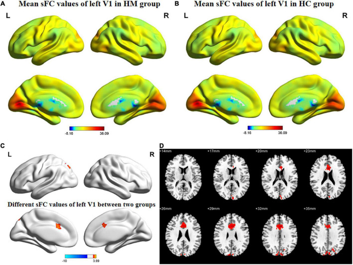
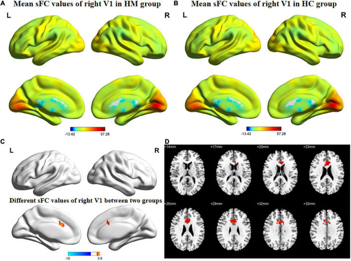
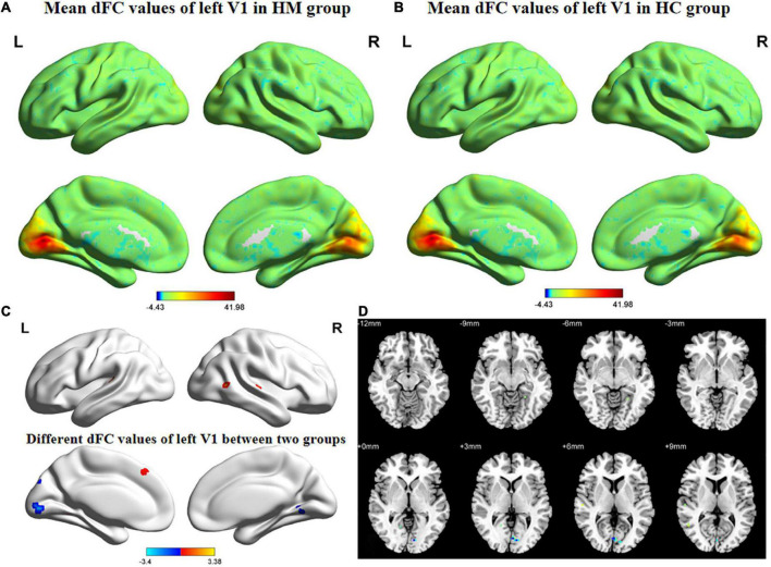
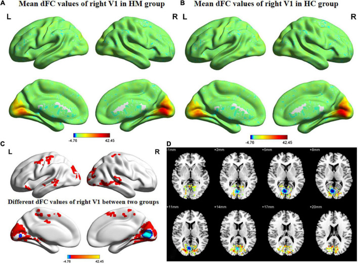
Results: In the left anterior cingulate gyrus (L-ACG)/left superior parietal gyrus (L-SPG) and left V1, sFC values were significantly greater in HM patients than in HCs. In the L-ACG and right V1, sFC values were also significantly greater in HM patients than in HCs [two-tailed, voxel-level P < 0.01, Gaussian random field (GRF) correction, cluster-level P < 0.05]. In the left calcarine cortex (L-CAL) and left V1, dFC values were significantly lower in HM patients than in HCs. In the right lingual gyrus (R-LING) and right V1, dFC values were also significantly lower in HM patients than in HCs (two-tailed, voxel-level P < 0.01, GRF correction, cluster-level P < 0.05).
Conclusion: Patients with HM exhibited significantly disturbed FC between the V1 and various brain regions, including L-ACG, L-SPG, L-CAL, and R-LING. This disturbance suggests that patients with HM could exhibit impaired cognitive and emotional processing functions, top-down control of visual attention, and visual information processing functions. HM patients and HCs could be distinguished from each other with high accuracy using sFC and dFC variabilities. These findings may help to identify the neural mechanism of decreased visual performance in HM patients.
Keywords: brain function; brain region; dynamic functional connectivity; high myopia; resting-state functional magnetic resonance imaging; seed-based functional connectivity analysis; static functional connectivity.
Copyright © 2023 Ji, Huang, Cheng, Fu, Zhong, Chen, Shu, Wei, Huang and Wu.
Conflict of interest statement
The authors declare that the research was conducted in the absence of any commercial or financial relationships that could be construed as a potential conflict of interest.
Figures
Similar articles
-
Exploration of Hippocampal Functional Connectivity Alterations in Patients with High Myopia via Seed-Based Functional Connectivity Analysis.Clin Ophthalmol. 2023 Nov 14;17:3443-3451. doi: 10.2147/OPTH.S434797. eCollection 2023. Clin Ophthalmol. 2023. PMID: 38026590 Free PMC article.
-
[Abnormal changes of static and dynamic functional connectivity of dopaminergic midbrain in patients with first-episode schizophrenia and their correlations with clinical symptoms].Zhonghua Yi Xue Za Zhi. 2023 Jun 6;103(21):1623-1630. doi: 10.3760/cma.j.cn112137-20221118-02428. Zhonghua Yi Xue Za Zhi. 2023. PMID: 37248062 Chinese.
-
Altered Functional Connectivity of the Primary Visual Cortex in Patients With Iridocyclitis and Assessment of Its Predictive Value Using Machine Learning.Front Immunol. 2021 May 7;12:660554. doi: 10.3389/fimmu.2021.660554. eCollection 2021. Front Immunol. 2021. PMID: 34025659 Free PMC article.
-
Alterations in degree centrality and functional connectivity in tension-type headache: a resting-state fMRI study.Brain Imaging Behav. 2024 Aug;18(4):819-829. doi: 10.1007/s11682-024-00875-w. Epub 2024 Mar 21. Brain Imaging Behav. 2024. PMID: 38512647 Review.
-
Classification and Prediction of Brain Disorders Using Functional Connectivity: Promising but Challenging.Front Neurosci. 2018 Aug 6;12:525. doi: 10.3389/fnins.2018.00525. eCollection 2018. Front Neurosci. 2018. PMID: 30127711 Free PMC article. Review.
Cited by
-
Single-cell RNA sequencing reveals anterograde trans-synaptic degeneration and exacerbated synaptic remodeling in myopia.Exp Mol Med. 2025 Jul;57(7):1536-1554. doi: 10.1038/s12276-025-01489-y. Epub 2025 Jul 3. Exp Mol Med. 2025. PMID: 40610752 Free PMC article.
-
Machine learning analysis reveals aberrant dynamic changes in amplitude of low-frequency fluctuations among patients with retinal detachment.Front Neurosci. 2023 Jul 20;17:1227081. doi: 10.3389/fnins.2023.1227081. eCollection 2023. Front Neurosci. 2023. PMID: 37547140 Free PMC article.
-
Exploration of Hippocampal Functional Connectivity Alterations in Patients with High Myopia via Seed-Based Functional Connectivity Analysis.Clin Ophthalmol. 2023 Nov 14;17:3443-3451. doi: 10.2147/OPTH.S434797. eCollection 2023. Clin Ophthalmol. 2023. PMID: 38026590 Free PMC article.
References
-
- Cheng Y., Chen X., Shi L., Li S., Huang H., Zhong P., et al. (2022). Abnormal functional connectivity between cerebral hemispheres in patients with high myopia: A Resting FMRI Study based on voxel-mirrored homotopic connectivity. Front. Hum. Neurosci. 16:910846. 10.3389/fnhum.2022.910846 - DOI - PMC - PubMed
-
- Corbetta M., Shulman G. (2002). Control of goal-directed and stimulus-driven attention in the brain. Nat. Rev. Neurosci. 3 201–215. - PubMed
LinkOut - more resources
Full Text Sources