

The value of convolutional neural networks-based deep learning model in differential diagnosis of space-occupying brain diseases
- PMID: 36816568
- PMCID: PMC9932812
- DOI: 10.3389/fneur.2023.1107957
The value of convolutional neural networks-based deep learning model in differential diagnosis of space-occupying brain diseases
Abstract
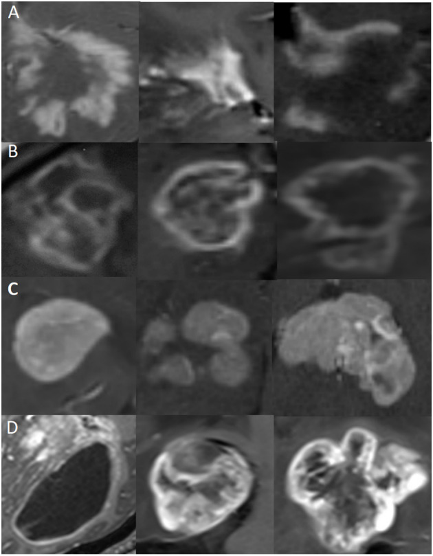
Objectives: It is still a challenge to differentiate space-occupying brain lesions such as tumefactive demyelinating lesions (TDLs), tumefactive primary angiitis of the central nervous system (TPACNS), primary central nervous system lymphoma (PCNSL), and brain gliomas. Convolutional neural networks (CNNs) have been used to analyze complex medical data and have proven transformative for image-based applications. It can quickly acquire diseases' radiographic features and correct doctors' diagnostic bias to improve diagnostic efficiency and accuracy. The study aimed to assess the value of CNN-based deep learning model in the differential diagnosis of space-occupying brain diseases on MRI.
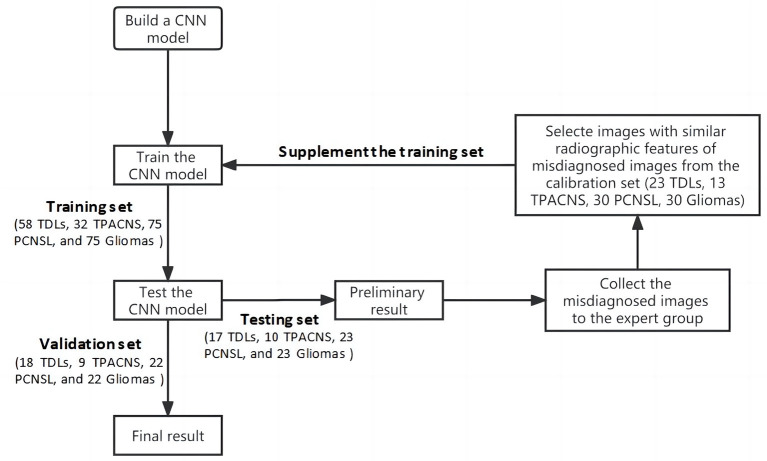
Methods: We retrospectively analyzed clinical and MRI data from 480 patients with TDLs (n = 116), TPACNS (n = 64), PCNSL (n = 150), and brain gliomas (n = 150). The patients were randomly assigned to training (n = 240), testing (n = 73), calibration (n = 96), and validation (n = 71) groups. And a CNN-implemented deep learning model guided by clinical experts was developed to identify the contrast-enhanced T1-weighted sequence lesions of these four diseases. We utilized accuracy, sensitivity, specificity, and area under the curve (AUC) to evaluate the performance of the CNN model. The model's performance was then compared to the neuroradiologists' diagnosis.
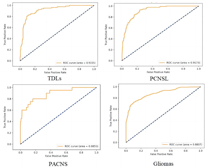
Results: The CNN model had a total accuracy of 87% which was higher than senior neuroradiologists (74%), and the AUC of TDLs, PCNSL, TPACNS and gliomas were 0.92, 0.92, 0.89 and 0.88, respectively.
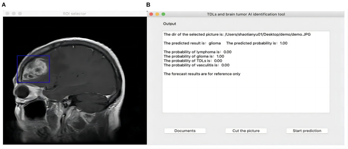
Conclusion: The CNN model can accurately identify specific radiographic features of TDLs, TPACNS, PCNSL, and gliomas. It has the potential to be an effective auxiliary diagnostic tool in the clinic, assisting inexperienced clinicians in reducing diagnostic bias and improving diagnostic efficiency.
Keywords: convolutional neural network; diagnosis; differential; magnetic resonance imaging; space-occupying brain lesions; tumefactive demyelinating lesions.
Copyright © 2023 Miao, Shao, Wang, Wang, Han, Li, Li, Sun, Wen and Liu.
Conflict of interest statement
The authors declare that the research was conducted in the absence of any commercial or financial relationships that could be construed as a potential conflict of interest.
Figures
Similar articles
-
Neuroimaging and clinicopathological differences between tumefactive demyelinating lesions and sentinel lesions of primary central nervous system lymphoma.Front Immunol. 2022 Aug 18;13:986473. doi: 10.3389/fimmu.2022.986473. eCollection 2022. Front Immunol. 2022. PMID: 36059526 Free PMC article.
-
Deep Learning for Automatic Differential Diagnosis of Primary Central Nervous System Lymphoma and Glioblastoma: Multi-Parametric Magnetic Resonance Imaging Based Convolutional Neural Network Model.J Magn Reson Imaging. 2021 Sep;54(3):880-887. doi: 10.1002/jmri.27592. Epub 2021 Mar 11. J Magn Reson Imaging. 2021. PMID: 33694250
-
Performance of Apparent Diffusion Coefficient Values and Conventional MRI Features in Differentiating Tumefactive Demyelinating Lesions From Primary Brain Neoplasms.AJR Am J Roentgenol. 2015 Nov;205(5):1075-85. doi: 10.2214/AJR.14.13970. AJR Am J Roentgenol. 2015. PMID: 26496556 Free PMC article.
-
Deep Learning With Data Enhancement for the Differentiation of Solitary and Multiple Cerebral Glioblastoma, Lymphoma, and Tumefactive Demyelinating Lesion.Front Oncol. 2021 Aug 18;11:665891. doi: 10.3389/fonc.2021.665891. eCollection 2021. Front Oncol. 2021. PMID: 34490082 Free PMC article.
-
Tumor-like Lesions in Primary Angiitis of the Central Nervous System: The Role of Magnetic Resonance Imaging in Differential Diagnosis.Diagnostics (Basel). 2024 Mar 14;14(6):618. doi: 10.3390/diagnostics14060618. Diagnostics (Basel). 2024. PMID: 38535038 Free PMC article. Review.
Cited by
-
Deep learning-driven modality imputation and subregion segmentation to enhance high-grade glioma grading.BMC Med Inform Decis Mak. 2025 May 30;25(1):200. doi: 10.1186/s12911-025-03029-0. BMC Med Inform Decis Mak. 2025. PMID: 40448035 Free PMC article.
References
LinkOut - more resources
Full Text Sources