. 2023 Jan 30:13:100120.
doi: 10.1016/j.ynpai.2023.100120. eCollection 2023 Jan-Jul.

Modulating the activity of human nociceptors with a SCN10A promoter-specific viral vector tool

Affiliations

Modulating the activity of human nociceptors with a SCN10A promoter-specific viral vector tool

Stephanie Mouchbahani-Constance et al. Neurobiol Pain. .

Abstract

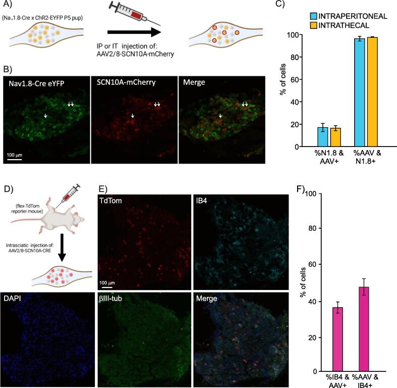
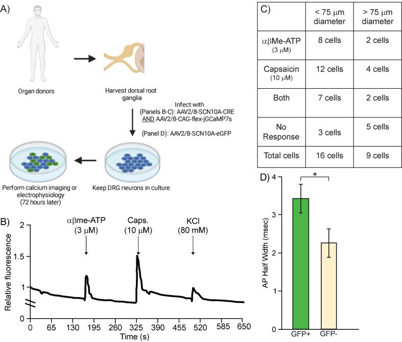
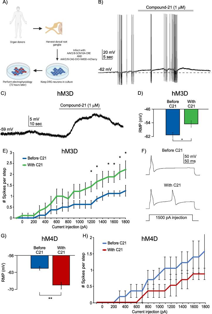
Despite the high prevalence of chronic pain as a disease in our society, there is a lack of effective treatment options for patients living with this condition. Gene therapies using recombinant AAVs are a direct method to selectively express genes of interest in target cells with the potential of, in the case of nociceptors, reducing neuronal firing in pain conditions. We designed a recombinant AAV vector expressing cargos whose expression was driven by a portion of the SCN10A (NaV1.8) promoter, which is predominantly active in nociceptors. We validated its specificity for nociceptors in mouse and human dorsal root ganglia and showed that it can drive the expression of functional proteins. Our viral vector and promoter package drove the expression of both excitatory or inhibitory DREADDs in primary human DRG cultures and in whole cell electrophysiology experiments, increased or decreased neuronal firing, respectively. Taken together, we present a novel viral tool that drives expression of cargo specifically in human nociceptors. This will allow for future specific studies of human nociceptor properties as well as pave the way for potential future gene therapies for chronic pain.

Keywords: AAV; Chemogenetics; DREADD; DRG; Gene therapy; Pain; Sodium channel.


Conflict of interest statement

The authors declare that they have no known competing financial interests or personal relationships that could have appeared to influence the work reported in this paper.

Figures

Fig. 1
Fig. 1
The AAV2/8-SCN10A-mCherry virus specifically infects DRG cells that express the NaV1.8 sodium channel, half of which seem to be non-peptidergic nociceptors. A) Schematic demonstrating expected results from injection of SCN10a-driven virus in NaV1.8-CrexChR2-EYFP P5 pups. B) Representative image of a DRG extracted from a NaV1.8-Cre × ChR2-eYFP pup injected with virus as described in panel A. eYFP-driven fluorescence from NaV1.8-Cre mouse (left) and mCherry-driven fluorescence driven by SCN10a virus (middle) with both images merged on the right. C) Quantification of proportion of eYFP (NaV1.8)-positive cells that are also mCherry (AAV)-positive (left 2 bars, 804/5151 and 858/5404 cells total, respectively), and quantification of proportion of mCherry (AAV)-positive cells that are also eYFP (NaV1.8)-positive (right 2 bars, 804/823 and 858/861 cells total, respectively). Both intraperitoneal and intrathecal injection routes were tested, both gave similar results. N = 3 mice for each the intraperitoneal and intrathecal injections. D) Schematic showing expected results from injection of SCN10a-Cre virus in flex-tdTom reporter mice. E) Immunohistochemistry of a DRG from a flex-TdTom reporter mouse injected intrasciatically with AAV2/8-SCN10A-Cre. TdTom fluorescence is driven by the virus’ expression, Isolectin-B4 was used to identify non-peptidergic nociceptors and βIII-tubulin was used to provide context along with DAPI. E) Quantification of proportion of IB4 neurons that are AAV (tdTom)-positive (left) (381/994 cells total from n = 3 mice) and proportion of AAV (tdTom)-positive neurons that are IB4-positive (381/806 total cells). Data are represented as Mean +/- SEM.
Fig. 2
Fig. 2
The virus AAV2/8-SCN10A-Cre successfully transduces human sensory neurons, which appear to be nociceptors, and drives the expression of functional GCaMP7s for calcium imaging. A) Schematic of experimental plan. B) Sample trace of calcium imaging using GCaMP7s, whose expression was driven by AAV2/8-SCN10a-Cre along with an AAV2/8-CAG-flex-GCaMP7s in human DRG neurons. Cells expressing GCaMP7s can successfully demonstrate a rise in intracellular calcium following applications of common algogens such as αβMe-ATP and Capsaicin. C) Quantification of number of infected human DRG neurons, classified based on the diameter of their soma, responding to αβMe-ATP, capsaicin, both or no response. n = 25 cells in total. D) Quantification of action potential half-width in human DRG neurons infected with an AAV2/8-SCN10a-GFP virus (n = 4 GFP-positive neurons) or that did not get infected with the virus (n = 5 GFP-negative neurons). * p < 0.05 (unpaired t-test).
Fig. 3
Fig. 3
Human DRG neuron excitability can be modulated by SCN10a-promoter-driven actuators in AAV2/8 viral constructs. A) Schematic of experimental plan. B) Sample trace of current clamp recording of a human DRG neuron infected by both AAV2/8-SCN10a-Cre and AAV2/8-CAG-DIO-hM3D-mCherry viruses. Application of Compound-21 (C21, agonist of hM3D) can increase firing rates of human DRG neurons infected with both viruses. C) Sample trace of gap-free current clamp recording of a human DRG neuron infected by hM3D dual virus approach and showing depolarization upon application of C21. D) Quantification of mean resting membrane potential (RMP) in mV of human DRG neurons infected with hM3D dual virus approach before C21 (blue bar) and after C21 (green bar). * p < 0.05, paired t-test. E) Input-output curve of human DRG neurons infected with hM3D dual virus approach before C21 (blue line, n = 8 neurons) and after C21 (green line, n = 8 neurons) following a current step protocol. * p < 0.05. Two-way ANOVA followed by the Tukey post hoc test. F) Representative trace of spikes in an m-Cherry-positive neuron before and after application of C21 in response to a 1500 pA step of current injection. G) Quantification of mean resting membrane potential (RMP) in mV of human DRG neurons infected with hM4D dual virus approach before C21 (blue bar) and after C21 (red bar). ** p < 0.021, paired t-test. H) Input-output curve of human DRG neurons infected with hM4D dual virus approach before C21 (blue line, n = 6 neurons) and after C21 (red line, n = 6 neurons) following a current step protocol. Two-way ANOVA followed by the Tukey post hoc test, no significant differences.

Similar articles

Cited by

References

    1. Asokan A., Schaffer D.V., Samulski R.J. The AAV vector toolkit: poised at the clinical crossroads. Mol. Ther. 2012;20:699–708. doi: 10.1038/mt.2011.287. - DOI - PMC - PubMed
    1. Bainbridge J.W.B., Smith A.J., Barker S.S., Robbie S., Henderson R., Balaggan K., Viswanathan A., Holder G.E., Stockman A., Tyler N., et al. Effect of gene therapy on visual function in Leber’s congenital amaurosis. N. Engl. J. Med. 2008;358:2231–2239. doi: 10.1056/NEJMoa0802268. - DOI - PubMed
    1. Bainbridge J.W.B., Mehat M.S., Sundaram V., Robbie S.J., Barker S.E., Ripamonti C., Georgiadis A., Mowat F.M., Beattie S.G., Gardner P.J., et al. Long-term effect of gene therapy on Leber’s congenital amaurosis. N. Engl. J. Med. 2015;372:1887–1897. doi: 10.1056/NEJMoa1414221. - DOI - PMC - PubMed
    1. Bennett J., Wellman J., Marshall K.A., McCague S., Ashtari M., DiStefano-Pappas J., Elci O.U., Chung D.C., Sun J., Wright J.F., et al. Safety and durability of effect of contralateral-eye administration of AAV2 gene therapy in patients with childhood-onset blindness caused by RPE65 mutations: a follow-on phase 1 trial. Lancet. 2016;388:661–672. doi: 10.1016/S0140-6736(16)30371-3. - DOI - PMC - PubMed
    1. Berta T., Qadri Y., Tan P.-H., Ji R.-R. Targeting dorsal root ganglia and primary sensory neurons for the treatment of chronic pain. Expert Opin. Ther. Targets. 2017;21:695–703. doi: 10.1080/14728222.2017.1328057. - DOI - PMC - PubMed

LinkOut - more resources