Review
. 2023 Feb 7;29(5):834-850.
doi: 10.3748/wjg.v29.i5.834.

Liver involvement in patients with COVID-19 infection: A comprehensive overview of diagnostic imaging features

Affiliations
Review

Liver involvement in patients with COVID-19 infection: A comprehensive overview of diagnostic imaging features

Davide Ippolito et al. World J Gastroenterol. .

Abstract

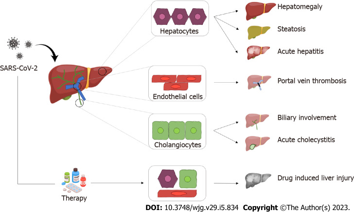
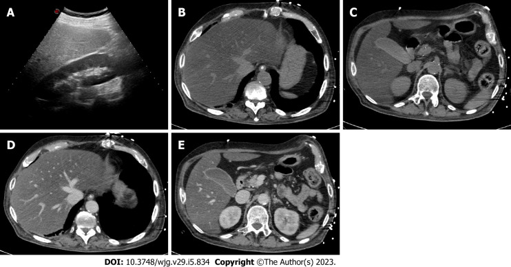
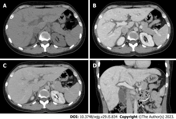
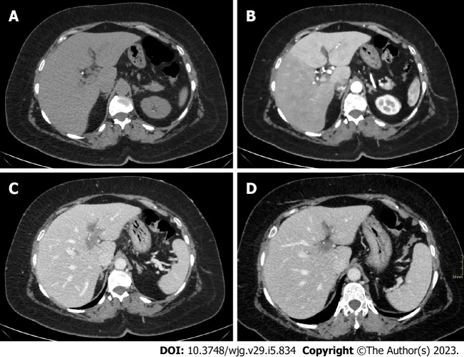
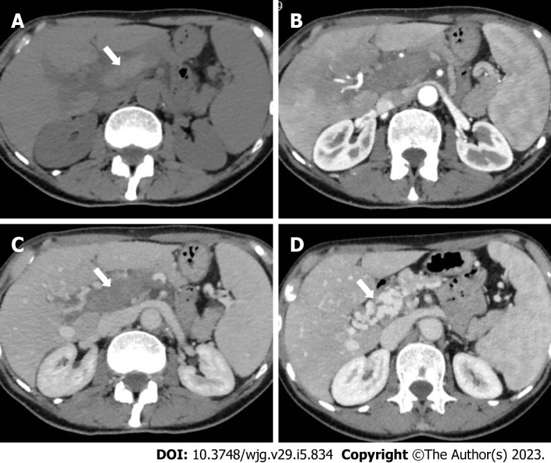
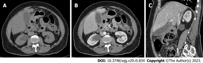
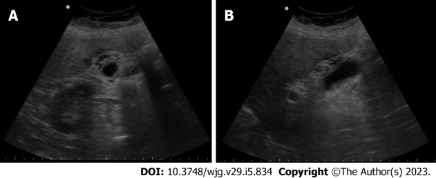
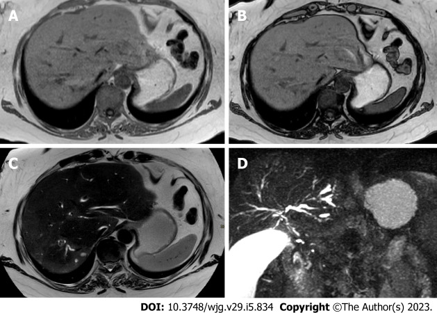
During the first wave of the pandemic, coronavirus disease 2019 (COVID-19) infection has been considered mainly as a pulmonary infection. However, different clinical and radiological manifestations were observed over time, including involvement of abdominal organs. Nowadays, the liver is considered one of the main affected abdominal organs. Hepatic involvement may be caused by either a direct damage by the virus or an indirect damage related to COVID-19 induced thrombosis or to the use of different drugs. After clinical assessment, radiology plays a key role in the evaluation of liver involvement. Ultrasonography (US), computed tomography (CT) and magnetic resonance imaging (MRI) may be used to evaluate liver involvement. US is widely available and it is considered the first-line technique to assess liver involvement in COVID-19 infection, in particular liver steatosis and portal-vein thrombosis. CT and MRI are used as second- and third-line techniques, respectively, considering their higher sensitivity and specificity compared to US for assessment of both parenchyma and vascularization. This review aims to the spectrum of COVID-19 liver involvement and the most common imaging features of COVID-19 liver damage.

Keywords: Adults; Biliary tract diseases; COVID-19; Fatty liver; Hepatic infarction; Hepatomegaly; Infection; Liver; Liver diseases; Liver failure; Magnetic resonance imaging; Pediatrics; SARS-CoV-2; Ultrasonography; X-Ray computed tomography.


Conflict of interest statement

Conflict-of-interest statement: Authors declare no conflict of interests for this article.

Figures

Figure 1
Figure 1
Graphical summary of the most common hepatic pathological findings in coronavirus disease 2019. SARS-CoV-2: Severe acute respiratory syndrome coronavirus 2.
Figure 2
Figure 2
A 45-year-old woman with coronavirus disease 2019 infection underwent abdominal ultrasonography due to elevated liver enzymes. A: As shown on ultrasonography, the liver is hyperechoic in comparison to the renal parenchyma, suggesting marked steatosis; B-E: The patient underwent contrast-enhanced computed tomography; B and C: On unenhanced images, the liver is diffusely hypoattenuating while its main vessels (i.e. portal vein and hepatic veins) appear brighter than the parenchyma due to a marked hepatic fatty infiltration; D and E: On post-contrast images, the liver is homogeneous without any focal lesion; C and E: The gallbladder is filled by sludge, with normal wall thickness. Finally, the combination of clinical and radiological findings allowed the final diagnosis of steatohepatitis.
Figure 3
Figure 3
A 33-year-old woman with coronavirus disease 2019 infection underwent contrast-enhanced computed tomography due to a suspicion of portal vein thrombosis. A: On unenhanced phase liver is enlarged, homogeneous and without any focal lesion; B: On the portal venous phase the liver enhancement is slight inhomogeneous associated with peri-portal edema, a typical finding of acute hepatitis; C and D: These aspects can be clearly observed also on the coronal reconstruction acquired on the portal venous phase (D) and the delayed phase (C).
Figure 4
Figure 4
A 40-year-old men with coronavirus disease 2019 infection and marked respiratory symptoms, underwent contrast-enhanced computed tomography due to elevated liver and biliary enzymes. A and B: On unenhanced phase (A) the liver is within normal limits. After the injection of contrast agent, on arterial phase (B) liver parenchyma shows inhomogeneous enhancement, with hypervascularization of the left liver lobe, as in case of transient hepatic attenuation differences; C: On the portal venous phase, the arterial hypervascularization fades to homogeneous enhancement and diffuse thrombosis of the left branch of the portal vein is demonstrated; D: After 6 mo the patient underwent contrast-enhanced computed tomography that demonstrated persistent portal vein thrombosis without venous collaterals.
Figure 5
Figure 5
A 62-year-old woman with acute portal vein thrombosis after coronavirus disease 2019 infection. A-C: Computed tomography (CT) images show acute thrombosis with hyperintense thrombus on unenhanced phase (A, arrow), heterogeneous enhancement of the liver parenchyma on hepatic arterial phase (B), and complete portal vein thrombosis on portal venous phase (C, arrow); D: Contrast-enhanced CT at 6-mo follow-up demonstrates chronic findings of portal cavernoma with multiple collateral vessels at the hepatic hilum (arrow).
Figure 6
Figure 6
A 66-year-old men with coronavirus disease 2019 infection, associated with abdominal pain in the upper quadrants. A-C: Contrast-enhanced computed tomography showed diffuse thickening of the gallbladder walls (A), with homogeneous contrast enhancement (B), better appreciable on the sagittal reconstruction (C). The gallbladder lumen is filled with biliary sludge (A) without calcified stones. The final diagnosis was acute acalculous cholecystitis.
Figure 7
Figure 7
A 53-year-old woman with coronavirus disease 2019 infection and markedly elevated biliary enzymes and C reactive protein levels. A and B: Ultrasonography demonstrates diffuse thickening of the gallbladder wall, associated with multiple small anechoic components into the walls, as in case of intramural abscesses. No stone is appreciated. The final diagnosis was acute acalculous cholecystitis.
Figure 8
Figure 8
A 44-year-old woman with coronavirus disease 2019 infection underwent magnetic resonance cholangiopancreatography due to elevated biliary enzymes with negative findings on ultrasonography and contrast-enhanced computed tomography. A and B: In-phase (A) and out-of-phase (B) imaging demonstrates hepatic homogeneous parenchyma without areas of fatty infiltration; C: On the T2-weighted image liver inflammation, especially in the right lobe, is characterized by areas of slight hyperenhancement, located nearby the biliary ducts; D: Finally, magnetic resonance cholangiopancreatography allows the evaluation of the biliary tree, showing multiple and diffuse focal biliary stenosis, in particular in the right lobe, with upstream dilation of small biliary ducts. This radiological aspect is typical of sclerosing cholangitis and the final diagnosis was a biliary involvement due to severe acute respiratory syndrome coronavirus 2.

Similar articles

Cited by

References

    1. World Health Organization. Health Emergency Dashboard. [cited 7 September 2022]. Available from: https://score.tools.who.int/tools/survey-populations-and-health-risks/to...
    1. Huang C, Wang Y, Li X, Ren L, Zhao J, Hu Y, Zhang L, Fan G, Xu J, Gu X, Cheng Z, Yu T, Xia J, Wei Y, Wu W, Xie X, Yin W, Li H, Liu M, Xiao Y, Gao H, Guo L, Xie J, Wang G, Jiang R, Gao Z, Jin Q, Wang J, Cao B. Clinical features of patients infected with 2019 novel coronavirus in Wuhan, China. Lancet. 2020;395:497–506. - PMC - PubMed
    1. Guan WJ, Ni ZY, Hu Y, Liang WH, Ou CQ, He JX, Liu L, Shan H, Lei CL, Hui DSC, Du B, Li LJ, Zeng G, Yuen KY, Chen RC, Tang CL, Wang T, Chen PY, Xiang J, Li SY, Wang JL, Liang ZJ, Peng YX, Wei L, Liu Y, Hu YH, Peng P, Wang JM, Liu JY, Chen Z, Li G, Zheng ZJ, Qiu SQ, Luo J, Ye CJ, Zhu SY, Zhong NS China Medical Treatment Expert Group for Covid-19. Clinical Characteristics of Coronavirus Disease 2019 in China. N Engl J Med. 2020;382:1708–1720. - PMC - PubMed
    1. Lechien JR, Chiesa-Estomba CM, De Siati DR, Horoi M, Le Bon SD, Rodriguez A, Dequanter D, Blecic S, El Afia F, Distinguin L, Chekkoury-Idrissi Y, Hans S, Delgado IL, Calvo-Henriquez C, Lavigne P, Falanga C, Barillari MR, Cammaroto G, Khalife M, Leich P, Souchay C, Rossi C, Journe F, Hsieh J, Edjlali M, Carlier R, Ris L, Lovato A, De Filippis C, Coppee F, Fakhry N, Ayad T, Saussez S. Olfactory and gustatory dysfunctions as a clinical presentation of mild-to-moderate forms of the coronavirus disease (COVID-19): a multicenter European study. Eur Arch Otorhinolaryngol. 2020;277:2251–2261. - PMC - PubMed
    1. Cummings MJ, Baldwin MR, Abrams D, Jacobson SD, Meyer BJ, Balough EM, Aaron JG, Claassen J, Rabbani LE, Hastie J, Hochman BR, Salazar-Schicchi J, Yip NH, Brodie D, O'Donnell MR. Epidemiology, clinical course, and outcomes of critically ill adults with COVID-19 in New York City: a prospective cohort study. Lancet. 2020;395:1763–1770. - PMC - PubMed