

Melanotic neuroectodermal tumor of infancy: A narrative review of dental aspects
- PMID: 36817027
- PMCID: PMC9931518
- DOI: 10.1016/j.sdentj.2022.12.003
Melanotic neuroectodermal tumor of infancy: A narrative review of dental aspects
Abstract
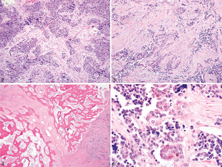
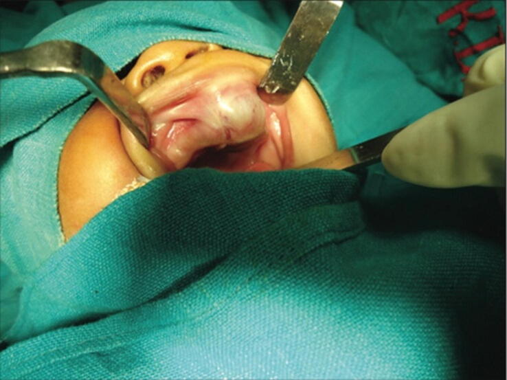
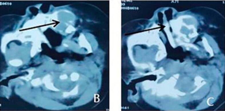
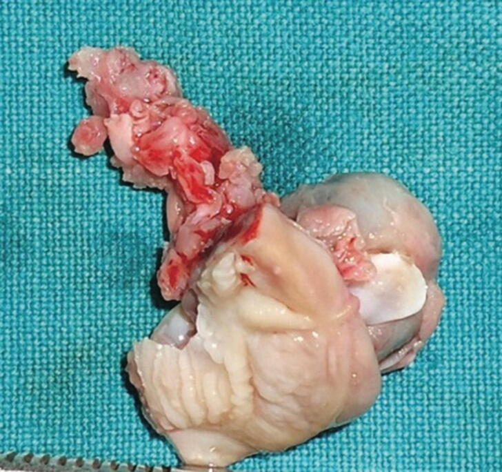
Melanotic neuroectodermal tumors of infancy (MNTI) are a rare type of benign tumor that affects the head and neck region. MNTI represents (68-80%) of the tumors in the maxillary region. This tumor is usually located in the alveolar crest, skull, mandible, and brain. Although this tumor is considered benign, it can grow rapidly, with a high risk of recurrence and interference with functions of infancy, such as feeding and breathing. It is also frequently harmful to the surrounding soft and hard tissues or adjacent sensitive vital structures. This study aimed to review the pathological, clinical presentation, and treatment of melanotic neuroectodermal tumors in infancy and the role of dentists in these cases.
Keywords: Complication; Dental; Infancy; MNTI; Treatment.
© 2022 The Authors.
Conflict of interest statement
The authors declare that they have no known competing financial interests or personal relationships that could have appeared to influence the work reported in this paper.
Figures
References
-
- Agrawal A., Joshi J., Agrawal D., Kumar P., Modi B. Oral melanotic neuroectodermal tumor of infancy: management of a case affecting the maxilla. J. Indian Soc. Pedodontics Prevent. Dent. 2020;38(3):319. - PubMed
-
- Almomani M.H., Rentea R.M. StatPearls Publishing; In StatPearls: 2022. Melanotic Neuroectodermal Tumor Of Infancy. - PubMed
-
- Chaudhary S., Manuja N., Ravishankar C.T., Sinha A., Vijayran M., Singh M. Oral melanotic neuroectodermal tumor of infancy. J. Indian Soc. Pedodontics Prevent. Dent. 2014;32(1):71. - PubMed
Publication types
LinkOut - more resources
Full Text Sources
