. 2023 Jan 19;25(3):90.
doi: 10.3892/ol.2023.13676. eCollection 2023 Mar.

HIF‑1α inhibits ferroptosis and promotes malignant progression in non‑small cell lung cancer by activating the Hippo‑YAP signalling pathway

Affiliations

HIF‑1α inhibits ferroptosis and promotes malignant progression in non‑small cell lung cancer by activating the Hippo‑YAP signalling pathway

Senzhong Zheng et al. Oncol Lett. .

Abstract

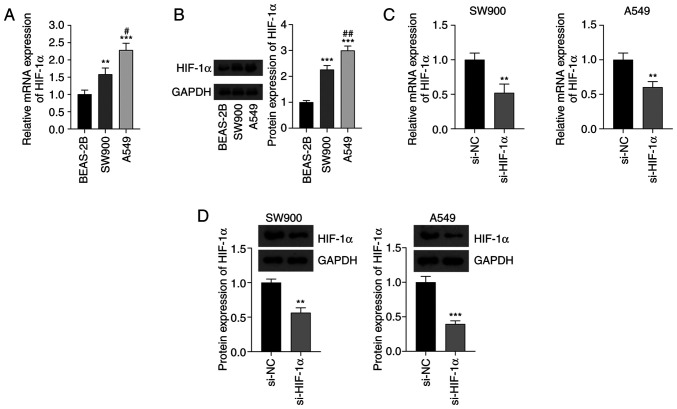
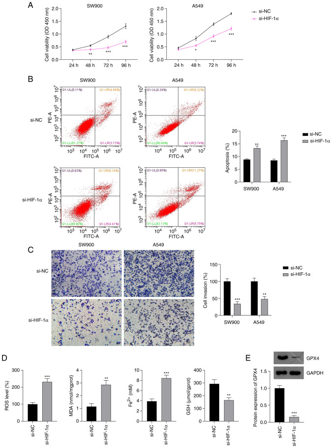
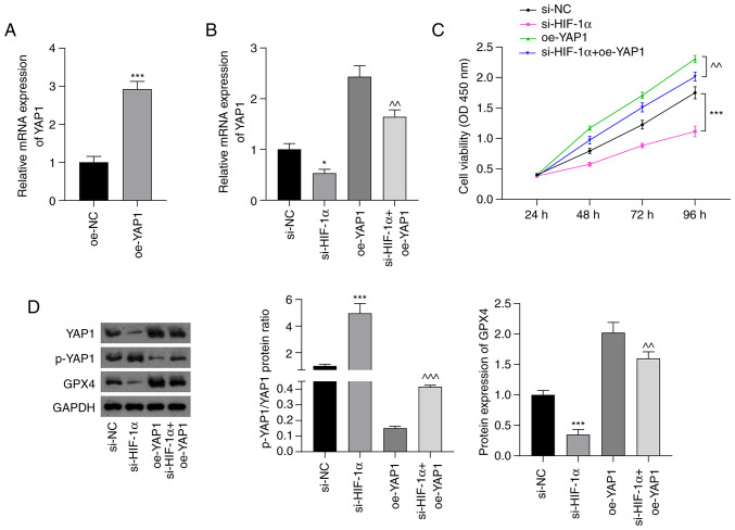
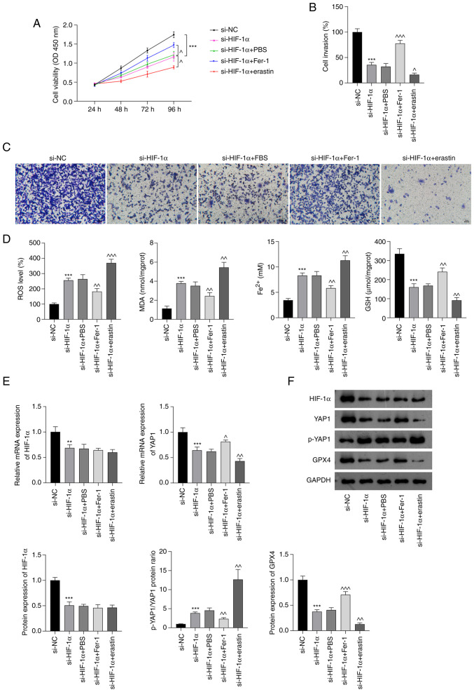
Ferroptosis and hypoxia-inducible factor 1α (HIF-1α) have critical roles in human tumors. The aim of the present study was to investigate the associations between ferroptosis, HIF-1α and cell growth in non-small cell lung cancer (NSCLC) cells. The lung cancer cell lines SW900 and A549 were evaluated using reverse transcription-quantitative polymerase chain reaction (RT-qPCR) to detect the expression of HIF-1α. Cell Counting Kit-8, flow cytometry and Transwell migration assays were used to measure cell viability, apoptosis and invasion, respectively. The production of reactive oxygen species (ROS) and levels of malondialdehyde (MDA), glutathione (GSH) and ferrous ion (Fe2+) were determined using detection kits. The expression levels of glutathione peroxidase 4 (GPX4) and Yes-associated protein 1 (YAP1) were detected using RT-qPCR and western blotting. The results showed that the expression of HIF-1α was significantly upregulated in NSCLC cells compared with normal human bronchial epithelial cells. Small interfering RNA specific to HIF-1α (si-HIF-1α) significantly decreased the proliferation and invasion of NSCLC cells and increased their apoptosis. si-HIF-1α also increased the levels of ROS, MDA and Fe2+ but decreased GSH and GPX4 levels in A549 cells. Additionally, si-HIF-1α increased phosphorylated (p-)YAP1 levels, suppressed GPX4 and YAP1 expression, and attenuated the YAP1 overexpression-induced changes in YAP1, p-YAP1 and GPX4 levels and cell viability. The ferroptosis antagonist ferrostatin-1 partially attenuated the effects of si-HIF-1α on the NSCLC cells, while the ferroptosis agonist erastin further inhibited NSCLC growth by blocking HIF-1α expression. In conclusion, the silencing of HIF-1α induces ferroptosis by suppressing Hippo-YAP pathway activation in NSCLC cells. The present study provides novel insights into the malignant progression of NSCLC and suggests that HIF-1α is an effective target for the treatment of NSCLC.

Keywords: Hippo-YAP pathway; ferroptosis; hypoxia-inducible factor 1α; non-small cell lung cancer; oxidative stress.


Conflict of interest statement

The authors declare that they have no competing interests.

Figures

Figure 1.
Figure 1.
Expression of HIF-1α in NSCLC cells. (A) Expression of HIF-1α mRNA in SW900 and A549 NSCLC cell lines and normal BEAS-2B cells determined using reverse transcription-quantitative polymerase chain reaction. (B) Results of the western blot analysis of HIF-1α protein. In (A) and (B), **P<0.01 and ***P<0.001 vs. BEAS-2B; and #P<0.05 and ##P<0.01 vs. SW900. (C) mRNA and (D) protein expression of HIF-1α in NSCLC cells transfected with si-HIF-1α. In (C) and (D), **P<0.01 and ***P<0.001 vs. si-NC. HIF-1α, hypoxia-inducible factor 1α; NSCLC, non-small cell lung cancer; si-HIF-1α, small interfering RNA to HIF-1α; si-NC, small interfering RNA negative control.
Figure 2.
Figure 2.
Effect of si-HIF-1α on the proliferation, invasion and ferroptosis of NSCLC cells. Results of (A) Cell Counting Kit-8, (B) flow cytometry and (C) Transwell invasion assays of NSCLC cells transfected with si-HIF-1α. (D) Levels of ROS, MDA, Fe2+ and GSH in A549 cells transfected with si-HIF-1α. Scale bar, 50 µm. (E) Protein expression of GPX4 in A549 cells transfected with si-HIF-1α. *P<0.05, **P<0.01 and ***P<0.001 vs. si-NC. si-HIF-1α, small interfering RNA to hypoxia-inducible factor 1α; NSCLC, non-small cell lung cancer; ROS, reactive oxygen species; MDA, malondialdehyde; Fe2+, ferrous iron; GSH, glutathione; GPX4, glutathione peroxidase 4; si-NC, small interfering RNA negative control; OD, optical density.
Figure 3.
Figure 3.
si-HIF-1α inhibits Hippo-YAP pathway activation in non-small cell lung cancer cells. (A) Relative mRNA expression of YAP1 in A549 cells transfected with oe-NC and oe-YAP1. ***P<0.001 vs. oe-NC. (B) Relative expression of YAP1 mRNA in cells transfected with si-HIF-1α and/or oe-YAP1. (C) Cell Counting Kit-8 assay results showing the viability of A549 cells. (D) Fold-change in the p-YAP1/YAP1 ratio and GPX4 protein expression assessed using western blot analysis. *P<0.05 and ***P<0.001 vs. si-NC; ^^P<0.01 and ^^^P<0.001 vs. oe-YAP1. si-HIF-1α, small interfering RNA to hypoxia-inducible factor 1 α; YAP, Yes-associated protein; p-, phosphorylated; oe-YAP1, overexpression of YAP1; oe-NC, overexpression negative control; si-NC, small interfering RNA negative control; GPX4, glutathione peroxidase 4; OD, optical density.
Figure 4.
Figure 4.
si-HIF-1α promotes ferroptosis by inhibiting Hippo-YAP pathway activation in non-small cell lung cancer cells. (A) Cell Counting Kit-8 assay results. (B) Quantified Transwell invasion assay results and (C) representative images of invaded A549 cells. Scale bar, 50 µm. (D) ROS, MDA, Fe2+ and GSH levels in A549 cells. (E) Relative mRNA expression of HIF-1α and YAP1 in A549 cells. (F) Protein levels of HIF-1α, p-YAP1/YAP1 and GPX4 in A549 cells. **P<0.01 and ***P<0.001 vs. si-NC. ^P<0.05, ^^P<0.01 and ^^^P<0.001 vs. si-HIF-1α. HIF-1α, hypoxia-inducible factor 1 α; si-HIF-1α, small interfering RNA to HIF-1α; YAP, Yes-associated protein; p-, phosphorylated; ROS, reactive oxygen species; MDA, malondialdehyde; Fe2+, ferrous iron; GSH, glutathione; GPX4, glutathione peroxidase 4; PBS, phosphate-buffered saline; Fer-1, ferrostatin-1; si-NC, small interfering RNA negative control; OD, optical density.

References

    1. Fashoyin-Aje LA, Fernandes LL, Sridhara R, Keegan P, Pazdur R. Demographic composition of lung cancer trials: FDA analysis. J Clin Oncol. 2018;36((15_suppl)):S9088. doi: 10.1200/JCO.2018.36.15_suppl.9088. - DOI
    1. Siegel RL, Miller KD, Jemal A. Cancer statistics, 2020. CA Cancer J Clin. 2020;70:7–30. doi: 10.3322/caac.21601. - DOI - PubMed
    1. Salgia R, Pharaon R, Mambetsariev I, Nam A, Sattler M. The improbable targeted therapy: KRAS as an emerging target in non-small cell lung cancer (NSCLC) Cell Rep Med. 2021;2:100186. doi: 10.1016/j.xcrm.2020.100186. - DOI - PMC - PubMed
    1. Goodgame B, Viswanathan A, Zoole J, Gao F, Miller CR, Subramanian J, Meyers BF, Patterson AG, Govindan R. Risk of recurrence of resected stage I non-small cell lung cancer in elderly patients as compared with younger patients. J Thorac Oncol. 2009;4:1370–1374. doi: 10.1097/JTO.0b013e3181b6bc1b. - DOI - PubMed
    1. Dixon SJ, Lemberg KM, Lamprecht MR, Skouta R, Zaitsev EM, Gleason CE, Patel DN, Bauer AJ, Cantley AM, Yang WS, et al. Ferroptosis: An iron-dependent form of nonapoptotic cell death. Cell. 2021;149:1060–1072. doi: 10.1016/j.cell.2012.03.042. - DOI - PMC - PubMed

LinkOut - more resources