. 2023 Feb 11:18:99-111.
doi: 10.2147/COPD.S391129. eCollection 2023.

Relationship Between ACSL4-Mediated Ferroptosis and Chronic Obstructive Pulmonary Disease

Affiliations

Relationship Between ACSL4-Mediated Ferroptosis and Chronic Obstructive Pulmonary Disease

Yingxi Wang et al. Int J Chron Obstruct Pulmon Dis. .

Abstract

Purpose: Although cigarette smoke exposure is the major risk factor for chronic obstructive pulmonary disease (COPD), the mechanism is not completely understood. The aim of the present study was to investigate whether ACSL4-mediated ferroptosis in lung epithelial cells plays a part in the COPD development process and its association.

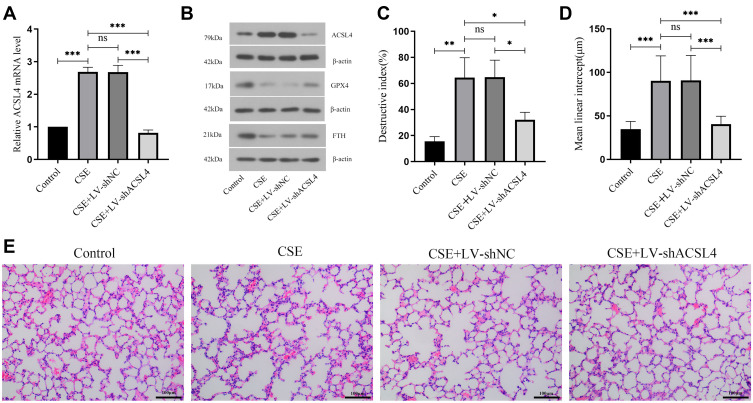
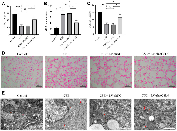
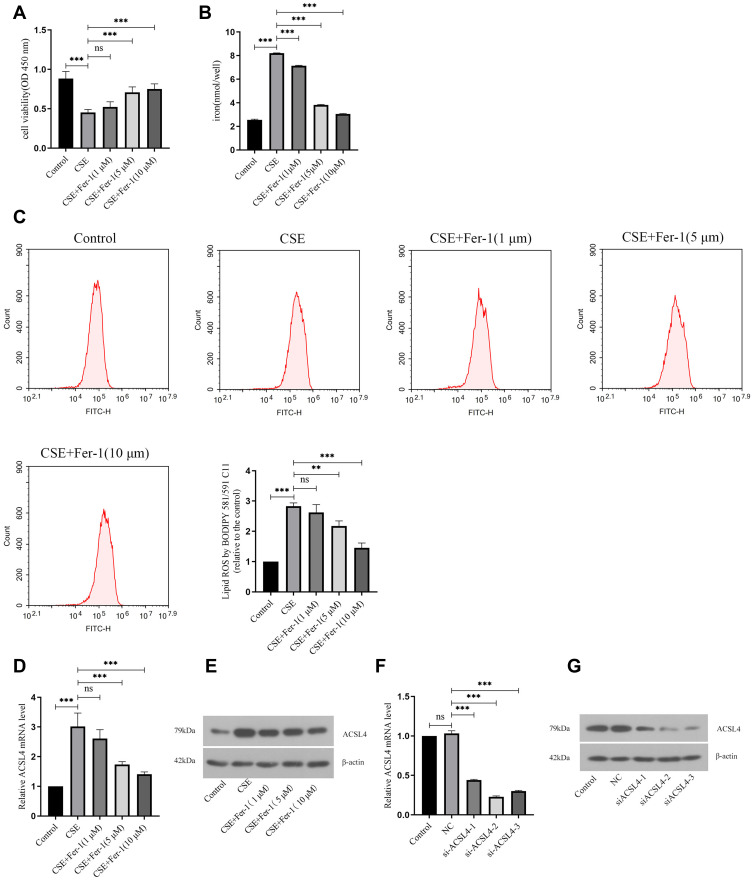
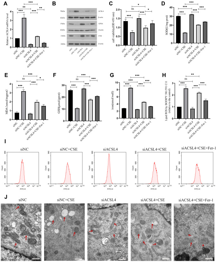
Patients and methods: In this study, animal and cell models of COPD were modelled using cigarette smoke extracts (CSEs), and cell viability, lipid ROS, iron ion deposition, and ferroptosis-related markers were measured in lung tissue and lung epithelial cells following CSE exposure. Morphological changes in mitochondria were observed in lung tissue and epithelial cells of the lung by transmission electron microscope. The expression levels of ACSL4 mRNA and protein in lung tissue and epithelial cells were measured by real-time PCR and Western blotting. In addition, animal-interfering lentivirus and cell-interfering RNA against ACSL4 were constructed in this study, ferroptosis in lung tissue and lung epithelial cells after ACSL4 interference was detected, and ACSL4 mRNA and protein expression levels were detected.

Results: CSE induced ferroptosis in lung tissues and lung epithelial cells, and the expression levels of ACSL4 were elevated in CSE-treated lung tissues and lung epithelial cells. After ACSL4 interference, the expression of ACSL4 decreased, mitochondrial morphology was restored, and ferroptosis in lung tissues and lung epithelial cells was alleviated. Both respiratory frequency and enhanced pause of COPD mice models decreased after ACSL4 interference.

Conclusion: ACSL4-mediated ferroptosis in lung epithelial cells is associated with COPD and positively correlated with ferroptosis in epithelial cells.

Keywords: ACSL4; chronic obstructive pulmonary disease; ferroptosis.


Conflict of interest statement

The authors declare no competing interests in this work.

Figures

Figure 1
Figure 1
ACSL4 is highly expressed in the lung tissue of COPD mice. (A) Relative expression of ACSL4 mRNA in mouse lung tissue by real-time PCR. (B) The expression of ACSL4 protein and ferroptosis-related proteins GPX4 and FTH in COPD mouse lung tissue was detected by Western blotting. (CE) HE staining of sections of mouse lung tissue. The DI and MLI values of each group were calculated. Data are presented as the mean ± SD of three replicates and analyzed using one-way ANOVA followed by post-hoc multiple comparisons. (nsp > 0.05, *p < 0.05, **p < 0.01, ***p < 0.001).
Figure 2
Figure 2
ACSL4 induces ferroptosis in lung tissue of COPD mice. (AC) Lung tissue from each group of mice was analysed for markers related to ferroptosis. (D) Iron deposition in lung tissue from mice in each group was observed by Prussian blue staining. (E) Transmission electron microscopy revealed the mitochondrial morphology in lung tissue from mice in each group; red arrows indicate mitochondria. Data are presented as the mean ± SD of three replicates and analyzed using one-way ANOVA followed by post-hoc multiple comparisons. (nsp > 0.05, *p < 0.05, **p < 0.01, ***p < 0.001).
Figure 3
Figure 3
ACSL4 induces ferroptosis in COPD lung epithelial cells. (A) CCK-8 determination of cell viability in each group. (B) Detection of iron ion content in cells of each group. (C) Lipid ROS content of cells in each group. (D) Relative expression levels of ACSL4 mRNA in each cell group. (E) ACSL4 protein expression in cells of each group. (F and G) Three siRNAs against human ACSL4 and control NC were constructed and transferred into BEAS-2B cells using transfection reagents. Real-time PCR and Western blotting were used to detect the ACSL4 mRNA and protein expression in the cells, respectively, and the siRNAs with the best interference efficiency were screened. Data are presented as the mean ± SD of three replicates and analyzed using one-way ANOVA followed by post-hoc multiple comparisons. (nsp > 0.05, **p < 0.01, ***p < 0.001).
Figure 4
Figure 4
Interference with ACSL4 can alleviate ferroptosis in lung epithelial cells. (A) Relative expression level of ACSL4 mRNA in each group of cells after siRNA interference. (B) ACSL4, GPX4 and FTH protein expression in each group after siRNA interference. (C) CCK-8 detection of cell viability in each group. (DF) Content detection of the ferroptosis related markers SOD, MDA and GSH. (G) Detection of iron ion content in cells of each group. (H and I) Detection of lipid ROS content by the BODIPY 581/591 C11 probe method. (J) Observation of mitochondrial morphology in cells of each group by transmission electron microscopy. Red arrows indicate mitochondria. Data are presented as the mean ± SD of three replicates and analyzed using one-way ANOVA followed by post-hoc multiple comparisons. (nsp > 0.05, *p < 0.05, **p < 0.01, ***p < 0.001).

References

    1. Nguyen JMK, Robinson DN, Sidhaye VK. Why new biology must be uncovered to advance therapeutic strategies for chronic obstructive pulmonary disease. Am J Physiol Lung Cell Mol Physiol. 2021;320(1):L1–L11. doi:10.1152/ajplung.00367.2020 - DOI - PMC - PubMed
    1. Sheikh K, Coxson HO, Parraga G. This is what COPD looks like. Respirology. 2016;21(2):224–236. doi:10.1111/resp.12611 - DOI - PubMed
    1. Sadeghi L, Yousefi Babadi V, Espanani HR. Toxic effects of the Fe2O3 nanoparticles on the liver and lung tissue. Bratisl Lek Listy. 2015;116(6):373–378. doi:10.4149/bll_2015_071 - DOI - PubMed
    1. Dixon SJ, Lemberg KM, Lamprecht MR, et al. Ferroptosis: an iron-dependent form of nonapoptotic cell death. Cell. 2012;149(5):1060–1072. doi:10.1016/j.cell.2012.03.042 - DOI - PMC - PubMed
    1. Yoshida M, Minagawa S, Araya J, et al. Involvement of cigarette smoke-induced epithelial cell ferroptosis in COPD pathogenesis. Nat Commun. 2019;10(1):3145. doi:10.1038/s41467-019-10991-7 - DOI - PMC - PubMed