

The transcriptional program during germinal center reaction - a close view at GC B cells, Tfh cells and Tfr cells
- PMID: 36817488
- PMCID: PMC9936310
- DOI: 10.3389/fimmu.2023.1125503
The transcriptional program during germinal center reaction - a close view at GC B cells, Tfh cells and Tfr cells
Abstract
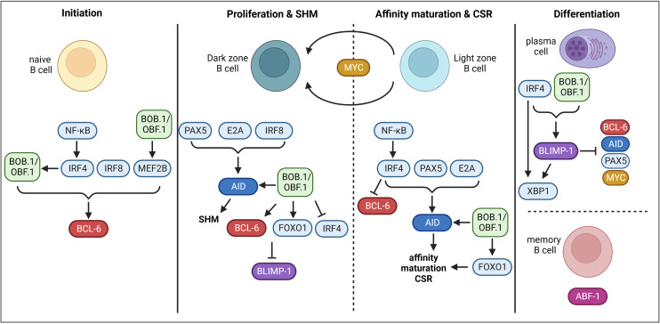
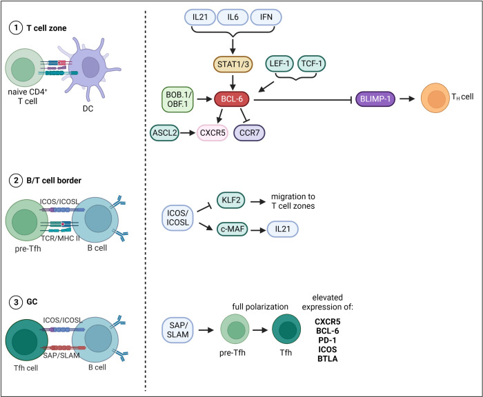
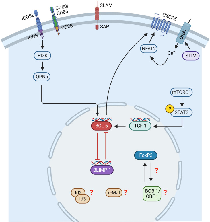
The germinal center (GC) reaction is a key process during an adaptive immune response to T cell specific antigens. GCs are specialized structures within secondary lymphoid organs, in which B cell proliferation, somatic hypermutation and antibody affinity maturation occur. As a result, high affinity antibody secreting plasma cells and memory B cells are generated. An effective GC response needs interaction between multiple cell types. Besides reticular cells and follicular dendritic cells, particularly B cells, T follicular helper (Tfh) cells as well as T follicular regulatory (Tfr) cells are a key player during the GC reaction. Whereas Tfh cells provide help to GC B cells in selection processes, Tfr cells, a specialized subset of regulatory T cells (Tregs), are able to suppress the GC reaction maintaining the balance between immune activation and tolerance. The formation and function of GCs is regulated by a complex network of signals and molecules at multiple levels. In this review, we highlight recent developments in GC biology by focusing on the transcriptional program regulating the GC reaction. This review focuses on the transcriptional co-activator BOB.1/OBF.1, whose important role for GC B, Tfh and Tfr cell differentiation became increasingly clear in recent years. Moreover, we outline how deregulation of the GC transcriptional program can drive lymphomagenesis.
Keywords: BOB.1/OBF.1; GC B cells; Tfh cells; Tfr cells; germinal center (GC); lymphoma; transcriptional regulation.
Copyright © 2023 Betzler, Ushmorov and Brunner.
Conflict of interest statement
The authors declare that the research was conducted in the absence of any commercial or financial relationships that could be construed as a potential conflict of interest.
Figures
Similar articles
-
Restoration of Follicular T Regulatory/Helper Cell Balance by OX40L-JAG1 Cotreatment Suppresses Lupus Nephritis in NZBWF1/j Mice.J Immunol. 2022 Jun 1;208(11):2467-2481. doi: 10.4049/jimmunol.2200057. Epub 2022 Apr 25. J Immunol. 2022. PMID: 35470257
-
Viral Replicative Capacity, Antigen Availability via Hematogenous Spread, and High TFH:TFR Ratios Drive Induction of Potent Neutralizing Antibody Responses.J Virol. 2019 Mar 5;93(6):e01795-18. doi: 10.1128/JVI.01795-18. Print 2019 Mar 15. J Virol. 2019. PMID: 30626686 Free PMC article.
-
Nonbinary Roles for T Follicular Helper Cells and T Follicular Regulatory Cells in the Germinal Center Response.J Immunol. 2023 Jul 1;211(1):15-22. doi: 10.4049/jimmunol.2200953. J Immunol. 2023. PMID: 37339403 Review.
-
Interleukin-21 promotes germinal center reaction by skewing the follicular regulatory T cell to follicular helper T cell balance in autoimmune BXD2 mice.Arthritis Rheumatol. 2014 Sep;66(9):2601-12. doi: 10.1002/art.38735. Arthritis Rheumatol. 2014. PMID: 24909430 Free PMC article.
-
Regulation of the Germinal Center Response.Front Immunol. 2018 Oct 25;9:2469. doi: 10.3389/fimmu.2018.02469. eCollection 2018. Front Immunol. 2018. PMID: 30410492 Free PMC article. Review.
Cited by
-
Novel Findings on the Development and Immunological Functions of Palatine Tonsils.Clin Rev Allergy Immunol. 2025 Jun 18;68(1):58. doi: 10.1007/s12016-025-09071-0. Clin Rev Allergy Immunol. 2025. PMID: 40531272 Review.
-
CD4+ T-cell-dependent differentiation of CD23+ follicular B cells contributes to the pulmonary pathology in a primary Sjögren's syndrome mouse model.Front Immunol. 2023 Jul 5;14:1217492. doi: 10.3389/fimmu.2023.1217492. eCollection 2023. Front Immunol. 2023. PMID: 37475871 Free PMC article.
-
Transcriptional Coactivator BOB1 (OBF1, OCA-B) Modulates the Specificity of DNA Recognition by the POU-Domain Factors OCT1 and OCT2 in a Monomeric Configuration.Biomolecules. 2024 Jan 17;14(1):123. doi: 10.3390/biom14010123. Biomolecules. 2024. PMID: 38254723 Free PMC article.
-
IL-2 Complex Therapy Mitigates Humoral Rejection of Fully Mismatched Skin Allografts by Inhibiting IgG Alloantibody Formation.Cells. 2025 Jul 16;14(14):1086. doi: 10.3390/cells14141086. Cells. 2025. PMID: 40710339 Free PMC article.
-
Accumulation of immune-suppressive CD4 + T cells in aging - tempering inflammaging at the expense of immunity.Semin Immunol. 2023 Nov;70:101836. doi: 10.1016/j.smim.2023.101836. Epub 2023 Aug 24. Semin Immunol. 2023. PMID: 37632992 Free PMC article. Review.
References
Publication types
MeSH terms
Substances
LinkOut - more resources
Full Text Sources
Research Materials
Miscellaneous