. 2023 Jan 31:14:1062211.
doi: 10.3389/fendo.2023.1062211. eCollection 2023.

Water deprivation induces hypoactivity in rats independently of oxytocin receptor signaling at the central amygdala

Affiliations

Water deprivation induces hypoactivity in rats independently of oxytocin receptor signaling at the central amygdala

Viviane Felintro et al. Front Endocrinol (Lausanne). .

Abstract

Introduction: Vasopressin (AVP) and oxytocin (OXT) are neuropeptides produced by magnocellular neurons (MCNs) of the hypothalamus and secreted through neurohypophysis to defend mammals against dehydration. It was recently demonstrated that MCNs also project to limbic structures, modulating several behavioral responses.

Methods and results: We found that 24 h of water deprivation (WD) or salt loading (SL) did not change exploration or anxiety-like behaviors in the elevated plus maze (EPM) test. However, rats deprived of water for 48 h showed reduced exploration of open field and the closed arms of EPM, indicating hypoactivity during night time. We evaluated mRNA expression of glutamate decarboxylase 1 (Gad1), vesicular glutamate transporter 2 (Slc17a6), AVP (Avpr1a) and OXT (Oxtr) receptors in the lateral habenula (LHb), basolateral (BLA) and central (CeA) amygdala after 48 h of WD or SL. WD, but not SL, increased Oxtr mRNA expression in the CeA. Bilateral pharmacological inhibition of OXTR function in the CeA with the OXTR antagonist L-371,257 was performed to evaluate its possible role in regulating the EPM exploration or water intake induced by WD. The blockade of OXTR in the CeA did not reverse the hypoactivity response in the EPM, nor did it change water intake induced in 48-h water-deprived rats.

Discussion: We found that WD modulates exploratory activity in rats, but this response is not mediated by oxytocin receptor signaling to the CeA, despite the upregulated Oxtr mRNA expression in that structure after WD for 48 h.

Keywords: central amygdala; dehydration; elevated plus maze test; exploratory behavior; oxytocin receptor.


Conflict of interest statement

The authors declare that the research was conducted in the absence of any commercial or financial relationships that could be construed as a potential conflict of interest.

Figures

Figure 1
Figure 1
Experimental procedures. Schematic illustration representing the experimental designs and basic procedures employed in the present study. “EPM”, elevated plus maze; “OF”, open field; “LHb”, Lateral habenula; “BLA”, basolateral amygdala; “CeA”, Central amygdala; “OXTR”, oxytocin receptor.
Figure 2
Figure 2
Effects of (A, B) 24 h or (C, D) 48 h of dehydration in male adult rats on (A, C) hematocrit and (B, D) plasma osmolality. Values are mean ± SD. The number of animals used per group was: Control = 9; 24 h WD = 11; 24 h SL = 10; 48 h WD = 11; 48 h SL = 9. Hematocrit data were submitted to one-way ANOVA followed by the Tukey post hoc test. Osmolality data were analyzed by the Kruskal-Wallis test followed by Dunn’s post hoc test. *p<0.05, **p<0.01 and ***p<0.001 compared to control groups; ##p<0.01 compared to the WD group.
Figure 3
Figure 3
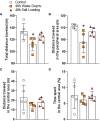
Effects of (A–F) 24 h or (G–L) 48 h of dehydration in male adult rats on (A, G) number of entries into closed arms, (B, H) percentage of entries into open arms, (C, I) percentage of time spent in open arms, (D, J) percentage of time spent in the central area, (E, K) number of head dipping episodes, and (F, L) number of stretch-attend postures during 5 min of evaluation in the elevated plus maze apparatus. Values are mean ± SD. The number of animals used per group was: 24 h Control = 11; 24 h WD = 10; 24 h SL = 10; 48 h Control = 12; 48 h WD = 12; 48 h SL = 12. Data were submitted to one-way ANOVA followed by the Tukey post hoc test, except for the percentage of time spent in the open arms after 24 h of dehydration and the number of stretch-attend postures after 48 h of dehydration, in which the Kruskal-Wallis test was used. **p<0.01 compared to control group; #p<0.05 compared to the WD group.
Figure 4
Figure 4
Effects of 48 h of dehydration in male adult rats on (A) total locomotion, (B) locomotion in the peripheral area, (C) locomotion in the central area, and (D) percentage the time spent in the central area during 10 min of evaluation in the open field test. Values are mean ± SD of 5 animals per group. Data were submitted to one-way ANOVA followed by the Tukey post hoc test, except for the distance traveled in the peripheral area, in which the Kruskal-Wallis test was used, followed by Dunn’s post hoc test. *p<0.05 compared to the control group.
Figure 5
Figure 5
Effects of 48 h of dehydration in male adult rats on relative mRNA expression of (A, E, I) the Vesicular Glutamate Transporter 2 (Slc17a6), (B, F, J) the Glutamate Decarboxylase 1 (Gad1), (C, G, K) the Arginine Vasopressin Receptor 1A (Avpr1a), and (D, H, L) the Oxytocin Receptor (Oxtr) in the (A–D) lateral habenula, (E–H) the basolateral amygdala (BLA), and (I-L) the central amygdala (CeA). Values are mean ± SD of 6 animals per group. Data of gene expression in BLA as well as Oxtr mRNA expression in CeA were analyzed by one-way ANOVA followed by the Tukey post hoc test. The other data were submitted to the Kruskal-Wallis test. *p<0.05 compared to the control group.
Figure 6
Figure 6
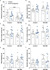
Effects of oxytocin receptor antagonist microinjection in the central amygdala of 48-h water-deprived male adult rats on (A) number of entries into closed arms, (B) percentage of entries into open arms, (C) percentage of time spent in open arms, (D) percentage of time spent in the central area, (E) number of head dipping episodes, and (F) number of stretch-attend postures during 5 min of evaluation in the elevated plus maze apparatus. Values are mean ± SD. The number of animals used per group was: Control + vehicle = 8; Control + antagonist = 10; 48 h WD + vehicle = 9; 48 h WD + antagonist = 9. Data were submitted to two-way ANOVA. The values of the number of stretch-attend postures were transformed to ranks before ANOVA. **p<0.01 comparing Water Deprived vs. Control groups.
Figure 7
Figure 7
Effects of oxytocin receptor antagonist microinjection in the central amygdala of 48 h water-deprived male adult rats on water intake during (A) 30 and (B) 120 minutes. Values are mean ± SD. The number of animals used per group was: Control + vehicle = 8; Control + antagonist = 10; 48 h WD + vehicle = 9; 48 h WD + antagonist = 9. Data were submitted to two-way ANOVA.

References

    1. Mecawi AS, Ruginsk SG, Elias LL, Varanda WA, Antunes-Rodrigues J. Neuroendocrine regulation of hydromineral homeostasis. Compr Physiol (2015) 5(3):1465–516. doi: 10.1002/cphy.c140031 - DOI - PubMed
    1. Knobloch HS, Charlet A, Hoffmann LC, Eliava M, Khrulev S, Cetin AH, et al. . Evoked axonal oxytocin release in the central amygdala attenuates fear response. Neuron (2012) 73(3):553–66. doi: 10.1016/j.neuron.2011.11.030 - DOI - PubMed
    1. Hernandez VS, Vazquez-Juarez E, Marquez MM, Jauregui-Huerta F, Barrio RA, Zhang L. Extra-neurohypophyseal axonal projections from individual vasopressin-containing magnocellular neurons in rat hypothalamus. Front Neuroanat (2015) 9:130. doi: 10.3389/fnana.2015.00130 - DOI - PMC - PubMed
    1. Zhang B, Qiu L, Xiao W, Ni H, Chen L, Wang F, et al. . Reconstruction of the hypothalamo-neurohypophysial system and functional dissection of magnocellular oxytocin neurons in the brain. Neuron (2021) 109(2):331–46.e7. doi: 10.1016/j.neuron.2020.10.032 - DOI - PubMed
    1. Hernandez VS, Hernandez OR, Perez de la Mora M, Gomora MJ, Fuxe K, Eiden LE, et al. . Hypothalamic vasopressinergic projections innervate central amygdala gabaergic neurons: Implications for anxiety and stress coping. Front Neural Circuits (2016) 10:92. doi: 10.3389/fncir.2016.00092 - DOI - PMC - PubMed

Publication types