. 2023 Feb 8:25:223-238.
doi: 10.1016/j.bioactmat.2023.02.004. eCollection 2023 Jul.

A "built-up" composite film with synergistic functionalities on Mg-2Zn-1Mn bioresorbable stents improves corrosion control effects and biocompatibility

Affiliations

A "built-up" composite film with synergistic functionalities on Mg-2Zn-1Mn bioresorbable stents improves corrosion control effects and biocompatibility

Zhenglong Dou et al. Bioact Mater. .

Abstract

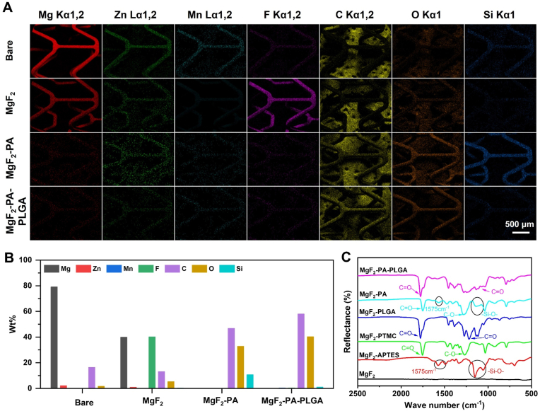
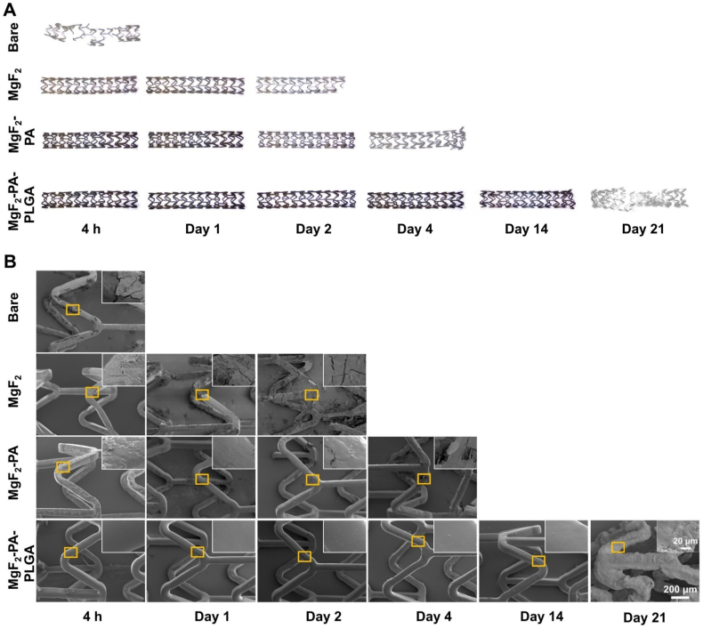
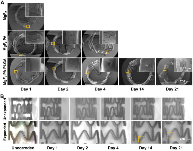
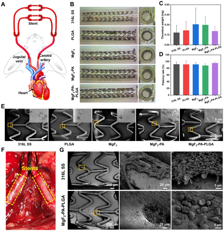
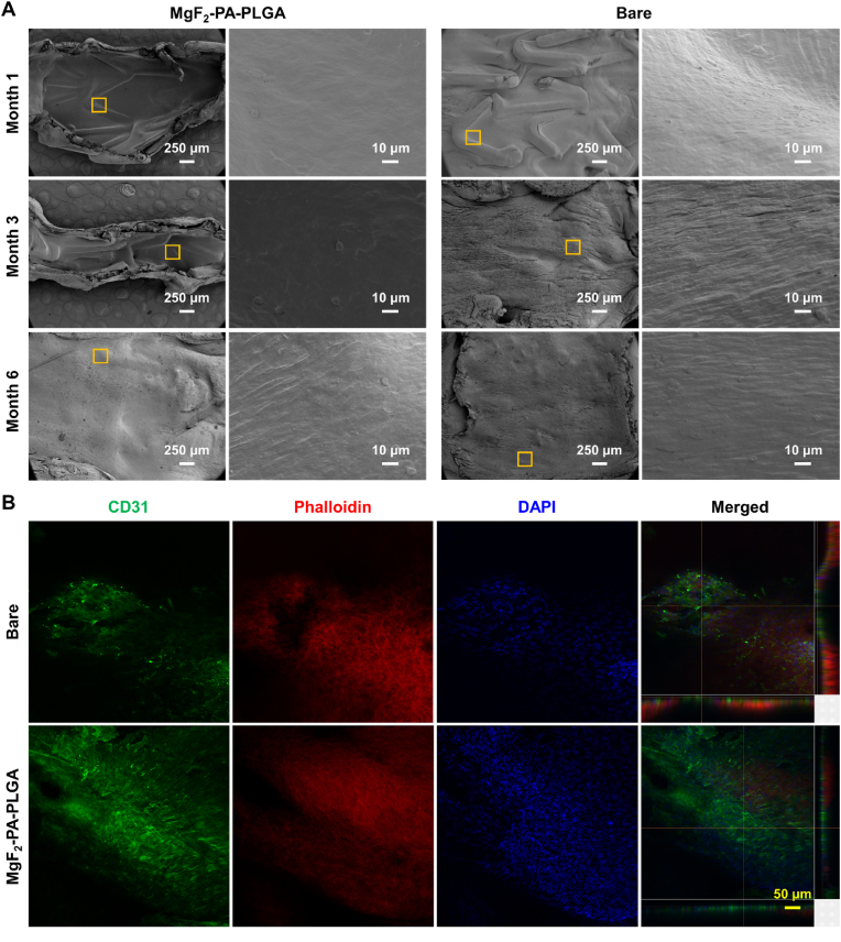
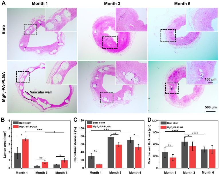
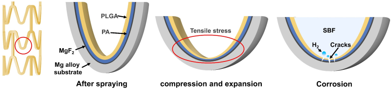
Control of premature corrosion of magnesium (Mg) alloy bioresorbable stents (BRS) is frequently achieved by the addition of rare earth elements. However, limited long-term experience with these elements causes concerns for clinical application and alternative methods of corrosion control are sought after. Herein, we report a "built-up" composite film consisting of a bottom layer of MgF2 conversion coating, a sandwich layer of a poly (1, 3-trimethylene carbonate) (PTMC) and 3-aminopropyl triethoxysilane (APTES) co-spray coating (PA) and on top a layer of poly (lactic-co-glycolic acid) (PLGA) ultrasonic spray coating to decorate the rare earth element-free Mg-2Zn-1Mn (ZM21) BRS for tailoring both corrosion resistance and biological functions. The developed "built-up" composite film shows synergistic functionalities, allowing the compression and expansion of the coated ZM21 BRS on an angioplasty balloon without cracking or peeling. Of special importance is that the synergistic corrosion control effects of the "built-up" composite film allow for maintaining the mechanical integrity of stents for up to 3 months, where complete biodegradation and no foreign matter residue were observed about half a year after implantation in rabbit iliac arteries. Moreover, the functionalized ZM21 BRS accomplished re-endothelialization within one month.

Keywords: Biocompatibility; Bioresorbable stents; Composite coating; Corrosion control; Magnesium alloys.


Conflict of interest statement

The authors declare that they have no known competing financial interests or personal relationships that could have appeared to influence the work reported in this paper.

Figures

Image 1
Graphical abstract
Fig. 1
Fig. 1
(A) Schematic diagram for preparing MgF2-PA-PLGA composite coating on ZM21 stents. (B) The surface morphology of the coatings after compression and expansion. (C) SEM images of strut cross-sections.
Fig. 2
Fig. 2
The surface elemental map (A) and element mass ratio (B) of the bare ZM21, MgF2, MgF2-PA, and MgF2-PA-PLGA stents obtained by EDS. (C) FT-IR diffuse reflection spectra of Mg samples coated with MgF2, MgF2-APTES, MgF2-PTMC, MgF2-PLGA, MgF2-PA, and MgF2-PA-PLGA.
Fig. 3
Fig. 3
In vitro corrosion evaluation of stents. (A) In vitro corrosion hydrogen collection device with ±0.05 ml resolution. (B) Hydrogen release curves after 96 h of immersion for Bare, PLGA, MgF2, MgF2-PA, and MgF2-PA-PLGA. (C) Hydrogen release behavior of the blank group and MgF2-PA-PLGA-coated stents immersed for 21 days. (D) Mg2+ concentration of MgF2-PA-PLGA stents at different immersion time points. (E) Mass loss of stents with MgF2-PA-PLGA coating that were immersed for 1, 2, 4, 7, 14, and 21 days. (F) Corrosion rates of Bare, PLGA, MgF2, MgF2-PA, and MgF2-PA-PLGA in vitro.
Fig. 4
Fig. 4
Corrosion morphologies of stents in vitro. (A) Macro morphology of stent after immersion at different time points. (b) SEM images of stents immersion at different time points. Insets are magnified images of the yellow rectangular area. (The empty area indicates that the stent has completely degraded.)
Fig. 5
Fig. 5
The cross-sectional photographs (A) and in situ corrosion morphologies (B) of stents after immersion at different times. (A) The cross-sectional SEM images of stent immersion for 1, 2, 4, 14, and 21 days. Insets are magnified images of the yellow rectangular area. (B) In situ corrosion morphologies of stents prior to corrosion, immersion for 1, 2, 4, 14, and 21 days. The area in the yellow dashed box indicates the occurrence of localized corrosion. (The empty area indicates that the stent has completely degraded.)
Fig. 6
Fig. 6
Radial support force of stents after immersion at different time points. (A) 1 day. (B) 2 days. (C) 4 days. (D) 7 days. (E) 14 days. (F) 21 days.
Fig. 7
Fig. 7
(A) Fluorescent staining of HUVECs cultured for 1 day, 3 days, and 5 days. (B) HUVECs' viability after 1 day, 3 days, and 5 days of incubation is calculated from CCK-8 tests. (C) Fluorescent staining of HUASMCs cultured for 1 day, 3 days, and 5 days. (D) HUASMCs' viability after 1 day, 3 days, and 5 days of incubation is calculated from CCK-8 tests. Data are presented as mean ± SD (n = 4), *p < 0.05, **p < 0.01, ***p < 0.001, ****p < 0.0001.
Fig. 8
Fig. 8
Semi-in vivo and in vivo hemocompatibility of stents. (A) A schematic diagram of semi-in vivo animal experiments. (B) Cross-section and appearance morphology after 2 h of blood circulation without additional heparin. (C) Thrombus mass after 2 h of blood circulation. (D) Tube patency rate after 2 h of blood circulation. (E) SEM images of the stent after blood circulation for 2 h. Insets are magnified images of the yellow rectangular area. (F) Image exhibiting the implantation of stents in the left and right iliac arteries. (G) SEM images of MgF2-PA-PLGA and 316L SS stents 2 h after in vivo implantation. Data are presented as mean ± SD (n = 4), *p < 0.05, **p < 0.01, ***p < 0.001, ****p < 0.0001.
Fig. 9
Fig. 9
Reconstructed 3D morphologies of MgF2-PA-PLGA stents after implantation at 1, 3, and 6 months by high-resolution micro-CT. (A-i, A-ii) One month after implantation, degradation occurred in some sites. (A-iii, A-iv) Support was lost 3 months after implantation due to severe deterioration. (A-v, A-vi) Completely degraded 6 months after implantation. (C) The volume of MgF2-PA-PLGA and corrosion products implanted at various times was calculated using Mimics Research 21.0 software. Changes of vascular wall thickness (D) and lumen area (E) at 1, 3, and 6 months of implantation. The data are presented as mean ± SD (n = 4), *p < 0.05, **p < 0.01, ***p < 0.001, ****p < 0.0001.
Fig. 10
Fig. 10
(A) SEM images revealed the degree of endothelialization of MgF2-PA-PLGA and bare ZM21 stents at 1 month, 3 months, and 6 months after implantation. (B) Confocal laser images to evaluate endothelialization 1 month after implantation of MgF2-PA-PLGA and bare ZM21 stents.
Fig. 11
Fig. 11
(A) Hematoxylin-eosin (H&E) staining images of iliac artery sections after 1, 3, and 6 months of implantation of MgF2-PA-PLGA and bare ZM21 stents. Vascular restenosis rate (B), lumen area (C), and vascular wall thickness (D) were calculated by Image J software. Data are presented as mean ± SD (n = 4), *p < 0.05, **p < 0.01, ***p < 0.001, ****p < 0.0001.
Fig. 12
Fig. 12
Schematic depicting the change of the stent's coating during expansion and the corrosion process during immersion.

Similar articles

Cited by

References

    1. Chen H., Yang W., Chen H., Liu L., Gao F., Yang X., Jiang Q., Zhang Q., Wang Y. Surface modification of mitoxantrone-loaded PLGA nanospheres with chitosan. Colloids Surf. B Biointerfaces. 2009;73(2):212–218. - PubMed
    1. Moravej M., Mantovani D. Biodegradable metals for cardiovascular stent application: interests and new opportunities. Int. J. Mol. Sci. 2011;12(7):4250–4270. - PMC - PubMed
    1. Heublein B., Rohde R., Kaese V., Niemeyer M., Hartung W., Haverich A. Biocorrosion of magnesium alloys: a new principle in cardiovascular implant technology? Heart. 2003;89(6):651–656. - PMC - PubMed
    1. Ding W. Opportunities and challenges for the biodegradable magnesium alloys as next-generation biomaterials. Regener. biomater. 2016;3(2):79–86. - PMC - PubMed
    1. Wu W., Chen S., Gastaldi D., Petrini L., Mantovani D., Yang K., Tan L., Migliavacca F. Experimental data confirm numerical modeling of the degradation process of magnesium alloys stents. Acta Biomater. 2013;9(10):8730–8739. - PubMed

LinkOut - more resources