

Editorial
doi: 10.26574/maedica.2022.17.4.999.
Extraarticular Synovial Osteochondromatosis in Baker's Cysts Bilaterally: a Rare Presentation
Affiliations
- PMID: 36818241
- PMCID: PMC9923057
- DOI: 10.26574/maedica.2022.17.4.999
Item in Clipboard
Editorial
Extraarticular Synovial Osteochondromatosis in Baker's Cysts Bilaterally: a Rare Presentation
Maedica (Bucur).
2022 Dec.
Abstract
Synovial osteochondromatosis is a rare benign pathology arising from the synovial membrane of the joints, synovial sheaths or uncommonly the bursae around the joints. Baker's cysts are fluid filled, synovium-lined lesions arising in popliteal fossa. Synovial chondromatosis involving the Baker`s cyst is extremely rare. The aim of this case report is to document this exceedingly rare extra articular synovial pathology involving Baker's cysts of bilateral knees and to emphasize the importance of radiographs and sonography in the diagnosis.
Figures

AP and lateral radiographs of bilateral knees showing soft tissue opacification (asterisks) in the posterior aspect of the joints with multiple calcified opacities (arrows) in the popliteal fossa
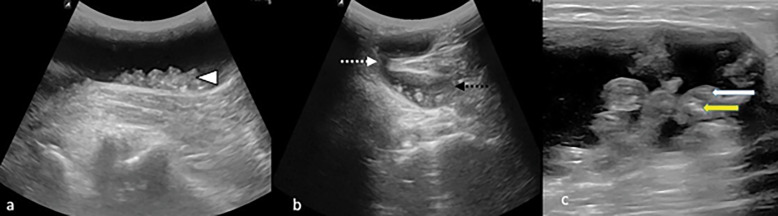
(a) Longitudinal sonogram of left popliteal fossa demonstrates cystic lesion with multiple intrabursal loose bodies (white arrow head); (b) transverse sonogram of right popliteal fossa showing base (black dotted arrow), neck (white dotted arrow) typical of Baker cyst with multiple loose bodies within the base; (c) magnified image of loose bodies shows hyperechoic center (yellow arrow) with hypoechoic chondroid periphery (white arrow)
References
-
- Milgram JW. Synovial osteochondromatosis: a histopathological study of thirty cases. J Bone Joint Surg. 1977;59:792. - PubMed
-
- Murphey MD, Vidal JA, Fanburg-Smith JC, Gajewski DA. Imaging of Synovial chondromatosis with radiologic-pathologic correlation. RadioGraphics. 2007;27:1465–1488. - PubMed
-
- Patel MR, Desai SS. Tenosynovial osteochondromatosis of the extensor tendon of a digit: case report and review of the literature. J Hand Surg. - PubMed
-
- Covall DJ, Fowble CD. Synovial chondromatosis of the biceps tendon sheath. Orthop Rev. 1994;23:902–905. - PubMed
Publication types
LinkOut - more resources
Full Text Sources