. 2023 Feb 2;3(1):vbad013.
doi: 10.1093/bioadv/vbad013. eCollection 2023.

An immune-suppressing protein in human endogenous retroviruses

Affiliations

An immune-suppressing protein in human endogenous retroviruses

Huan Zhang et al. Bioinform Adv. .

Abstract

Motivation: Retroviruses are important contributors to disease and evolution in vertebrates. Sometimes, retrovirus DNA is heritably inserted in a vertebrate genome: an endogenous retrovirus (ERV). Vertebrate genomes have many such virus-derived fragments, usually with mutations disabling their original functions.

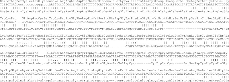
Results: Some primate ERVs appear to encode an overlooked protein. This protein is homologous to protein MC132 from Molluscum contagiosum virus, which is a human poxvirus, not a retrovirus. MC132 suppresses the immune system by targeting NF- κ B, and it had no known homologs until now. The ERV homologs of MC132 in the human genome are mostly disrupted by mutations, but there is an intact copy on chromosome 4. We found homologs of MC132 in ERVs of apes, monkeys and bushbaby, but not tarsiers, lemurs or non-primates. This suggests that some primate retroviruses had, or have, an extra immune-suppressing protein, which underwent horizontal genetic transfer between unrelated viruses.

Contact: mcfrith@edu.k.u-tokyo.ac.jp.


Figures

Fig. 1.
Fig. 1.
Alignment between Dfam’s HERV30 consensus DNA sequence and MC132 protein (Q98298). The DNA’s translation is shown above it. ‖| indicates a match, ::: a positive substitution score, and … a zero substitution score (Fig. 7). The alignment has 36% identity. Lowercase regions were deemed to be simple repeats by the alignment tool (LAST)
Fig. 2.
Fig. 2.
Location of the newly-discovered protein in an ERV in human chromosome 4. The location of the new protein is shown by the top bar labeled ‘refiner’. The black bars below that show DNA segments aligned to known transposable element proteins [from a previous study (Frith, 2022)], which are in the usual gag-pol-env order. Below that are RepeatMasker annotations of transposable element-derived segments. Here, RepeatMasker annotates two long terminal repeats of type LTR30, flanking an internal retroviral sequence of type HERV30. There is a 3881-bp deletion near the end of this internal sequence. Screenshot from the UCSC genome browser (http://genome.ucsc.edu) (Kent et al., 2002)
Fig. 3.
Fig. 3.
The new protein overlaps gaps in HERV17 annotations. The panels show three human genome locations with homology to the new protein (bars labeled ‘refiner’). The protein aligns to each location as two or three separate fragments. The black bars below that show DNA segments aligned to known transposable element proteins. Below that are RepeatMasker annotations of transposable element-derived segments. In each case, the new protein overlaps a retroviral sequence of type HERV17. However, in each case, the protein overlaps a consistent gap in RepeatMasker’s HERV17 annotation. There is also an unexpected pol protein homology (HERVIP10F_pol) between the new protein and the gag gene
Fig. 4.
Fig. 4.
Locations of the newly-found protein in four ERV families. The right-hand panel shows the full-length ERVs, and the left-hand panel is zoomed in to the matching region. HERV30 has a long match to the protein (green), while the other ERVs have fragmentary matches. The separated matches in HERV17 (red) appear to correspond to the annotation gap shown in Figure 3
Fig. 5.
Fig. 5.
The new protein matches a gap in ERV annotation in the bushbaby genome. The black bar shows a genome segment homologous to MC132, and the gray bars show RepeatMasker annotations. The colored bars show alignments to human and tarsier genomes: these alignments do not cover the segment homologous to MC132
Fig. 6.
Fig. 6.
Evolutionary tree of protein fossils homologous to MC132. Blue indicates fossils from Platyrrhini (new-world monkey) genomes, red Catarrhini (apes and old-world monkeys), and green bushbaby. Pink circles mark branches with medium-to-high confidence (bootstrap value >70%)
Fig. 7.
Fig. 7.
The substitution score matrix inferred by last-train

References

    1. Baertsch R. et al. (2008) Retrocopy contributions to the evolution of the human genome. BMC Genomics, 9, 1–19. - PMC - PubMed
    1. Brady G. et al. (2015) Poxvirus protein MC132 from molluscum contagiosum virus inhibits NF-κB activation by targeting p65 for degradation. J. Virol., 89, 8406–8415. - PMC - PubMed
    1. Frith M.C. (2022) Paleozoic protein fossils illuminate the evolution of vertebrate genomes and transposable elements. Mol. Biol. Evol., 39, msac068. - PMC - PubMed
    1. Gouveia-Oliveira R. et al. (2007) MaxAlign: maximizing usable data in an alignment. BMC Bioinformatics, 8, 1–8. - PMC - PubMed
    1. Grandi N. et al. (2016) Contribution of type W human endogenous retroviruses to the human genome: characterization of HERV-W proviral insertions and processed pseudogenes. Retrovirology, 13, 1–25. - PMC - PubMed