. 2023 Feb 3:14:1072168.
doi: 10.3389/fpls.2023.1072168. eCollection 2023.

Phylogenetic and AlphaFold predicted structure analyses provide insights for A1 aspartic protease family classification in Arabidopsis

Affiliations

Phylogenetic and AlphaFold predicted structure analyses provide insights for A1 aspartic protease family classification in Arabidopsis

Yanling Duan et al. Front Plant Sci. .

Abstract

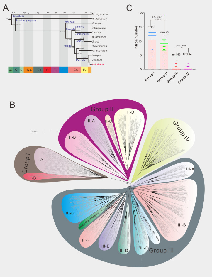
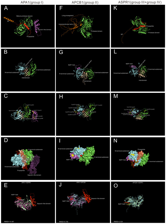
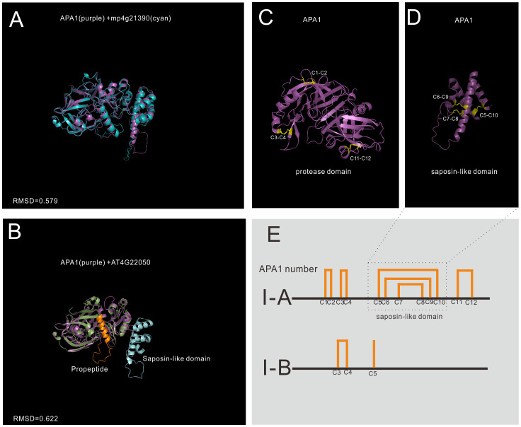
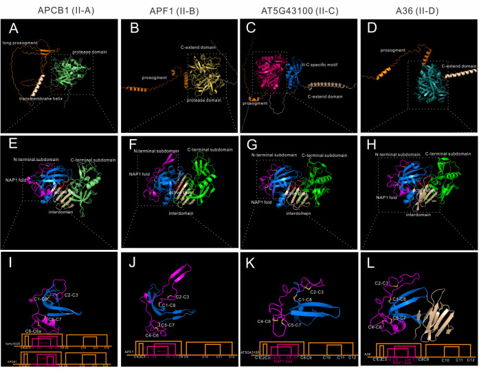
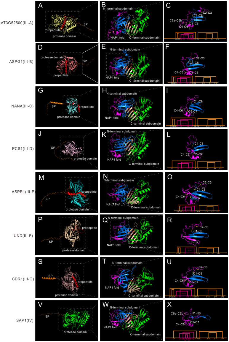
Aspartic proteases are widely distributed in animals, plants, fungi and other organisms. In land plants, A1 aspartic protease family members have been implicated to play important and varied roles in growth, development and defense. Thus a robust classification of this family is important for understanding their gene function and evolution. However, current A1 family members in Arabidopsis are less well classified and need to be re-evaluated. In this paper, 70 A1 aspartic proteases in Arabidopsis are divided into four groups (group I-IV) based on phylogenetic and gene structure analyses of 1200 A1 aspartic proteases which are obtained from 12 Embryophyta species. Group I-III members are further classified into 2, 4 and 7 subgroups based on the AlphaFold predicted structures. Furthermore, unique insights of A1 aspartic proteases have been unraveled by AlphaFold predicted structures. For example, subgroup II-C members have a unique II-C specific motif in the C-extend domain, and subgroup IV is a Spermatophyta conserved group without canonical DTGS/DSGT active sites. These results prove that AlphaFold combining phylogenetic analysis is a promising solution for complex gene family classification.

Keywords: AlphaFold; Arabidopsis thaliana; aspartic protease; cysteine residues; phylogentic analysis.


Conflict of interest statement

The authors declare that the research was conducted in the absence of any commercial or financial relationships that could be construed as a potential conflict of interest.

Figures

Figure 1
Figure 1
Phylogenetic analysis of A1 aspartic protease family in 12 selected species. (A) The evolution relationship of 12 selected species. The top of tree showed the absolute age, unit: million years, the bottom of the tree showed the geologic time, C, Cambrian; O, Ordovician; S, Silurian; De, Devonian; Ca, Carboniferous; P, Permian; T, Triassic; Ju, Jurassic; Cr, Cretaceous; P, Paleogene (from left to right). (B) Phylogenetic tree of A1 family members in 12 selected species, maximum likelihood method (ML) was applied with a 1000 bootstrap analysis, groups and subgroups are labeled. (C) Average intron numbers of group I-IV, n presents gene numbers in each group.
Figure 2
Figure 2
AlphaFold predicted structure models of Group I-IV. (A–D) Group I typical structure (APA1). (A) overall structure of APA1. SP,signal peptide, is colored by orange; propeptide is colored by red, saposin-like domain is colored by purple. (B) Proposed mature protease domain of APA1. N-terminal subdomain is colored by cyan, C-terminal subdomain is colored by green, interdomain beta-sheet is colored by wheat, and active sites are colored by red. (C) Disulfide bonds of APA1. (D) Surface and mesh of APA. The proposed mature protease structure is presented by surface, propeptide and saposin-like domain is presented by mesh. (F–I) Group II typical structure (APCB1). (F) Overall structure of APCB1.Long prosegment is colored by orange and transmembrane helix is colored by wheat. (B) Proposed protease domain of APCB1,NAP1 fold is clored by purple. (H) Disulfide bonds of APCB1. (I) surface of APCB1. (K–N) Group III typical structure (ASPR1). (K) Overall structure of APSR1. (L) proposed mature protease structure of ASPR1. (M) Surface of ASPR1. (N) Disulfide bonds of ASPR1. (E–O) Superposition analysis of APA1, APCB1 and ASPR1, RMSD value are presented.
Figure 3
Figure 3
AlphaFold predicted structure models of group I (subgroup I-A and subgroup II-B) (A) Structure alignment of APA1 and its Marchantia polymorpha mp4g21390; (B) Structure alignment of subgroup I-A (APA1) and subgroup I-B (AT4G22050); (C) The disulfide bonds of mature APA1 protease domain; (D) The disulfide bonds of saposin-like domain; (E) Disulfide bond topology of subgroup I-A and I-B.
Figure 4
Figure 4
AlphaFold predicted structure models of group II (subgroup II-A to II-D). (A–D) Overall structures of APCB1, APF1, AT5G43100 and A36. (E–H) Protease domain of APCB1, APF1, AT5G43100 and A36. (I–L) NAP1 fold structure and disulfide bond topology of APCB1, APF1, AT5G43100 and A36.
Figure 5
Figure 5
AlphaFold predicted structure models of group III and group IV. (A-C) Structure and disulfide bond topology of III-B(AT3G52500); (D–F) Structure and disulfide bond topology of III-C (ASPG1); (G–I) Structure and disulfide bond topology of III-D (NANA); (J–L) Structure and disulfide bond topology of III-E (PCS1); (M–O) Structure and disulfide bond topology of III-F (ASPR1); (P–R) Structure and disulfide bond topology of III-G (UND); (S–U) Structure and disulfide bond topology of III-H (CDR1); (V–X). Structure and disulfide bond topology of IV (SAP1).

Similar articles

Cited by

References

    1. Athauda S. B., Matsumoto K., Rajapakshe S., Kuribayashi M., Kojima M., Kubomura-Yoshida N., et al. . (2004). Enzymic and structural characterization of nepenthesin, a unique member of a novel subfamily of aspartic proteinases. Biochem. J. 381, 295–306. doi: 10.1042/BJ20031575 - DOI - PMC - PubMed
    1. Beers E. P., Jones A. M., Dickerman A. W. (2004). The S8 serine, C1A cysteine and A1 aspartic protease families in. Arabidopsis. Phytochemistry. 65, 43–58. doi: 10.1016/j.phytochem.2003.09.005 - DOI - PubMed
    1. Breitenbach H. H., Wenig M., Wittek F., Jordá L., Maldonado-Alconada A. M., Sarioglu H., et al. . (2014). Contrasting roles of the apoplastic aspartyl protease APOPLASTIC, ENHANCED DISEASE SUSCEPTIBILITY1-DEPENDENT1 and LEGUME LECTIN-LIKE PROTEIN1 in Arabidopsis systemic acquired resistance. Plant Physiol. 165, 791–809. doi: 10.1104/pp.114.239665 - DOI - PMC - PubMed
    1. Cao S., Guo M., Wang C., Xu W., Shi T., Tong G., et al. . (2019). Genome-wide characterization of aspartic protease (AP) gene family in populus trichocarpa and identification of the potential PtAPs involved in wood formation. BMC Plant Biol. 19, 276. doi: 10.1186/s12870-019-1865-0 - DOI - PMC - PubMed
    1. Chen J., Ouyang Y., Wang L., Xie W., Zhang Q. (2009). Aspartic proteases gene family in rice: Gene structure and expression, predicted protein features and phylogenetic relation. Gene 442, 108–118. doi: 10.1016/j.gene.2009.04.021 - DOI - PubMed

LinkOut - more resources