. 2023 Feb 1;13(2):1126-1137.
doi: 10.21037/qims-22-565. Epub 2023 Jan 5.

A magnetic resonance imaging-based computational analysis of cerebral hemodynamics in patients with carotid artery stenosis

Affiliations

A magnetic resonance imaging-based computational analysis of cerebral hemodynamics in patients with carotid artery stenosis

Jonas Schollenberger et al. Quant Imaging Med Surg. .

Abstract

Management of asymptomatic carotid artery stenosis (CAS) relies on measuring the percentage of stenosis. The aim of this study was to investigate the impact of CAS on cerebral hemodynamics using magnetic resonance imaging (MRI)-informed computational fluid dynamics (CFD) and to provide novel hemodynamic metrics that may improve the understanding of stroke risk. CFD analysis was performed in two patients with similar degrees of asymptomatic high-grade CAS. Three-dimensional anatomical-based computational models of cervical and cerebral blood flow were constructed and calibrated patient-specifically using phase-contrast MRI flow and arterial spin labeling perfusion data. Differences in cerebral hemodynamics were assessed in preoperative and postoperative models. Preoperatively, patient 1 demonstrated large flow and pressure reductions in the stenosed internal carotid artery, while patient 2 demonstrated only minor reductions. Patient 1 exhibited a large amount of flow compensation between hemispheres (80.31%), whereas patient 2 exhibited only a small amount of collateral flow (20.05%). There were significant differences in the mean pressure gradient over the stenosis between patients preoperatively (26.3 vs. 1.8 mmHg). Carotid endarterectomy resulted in only minor hemodynamic changes in patient 2. MRI-informed CFD analysis of two patients with similar clinical classifications of stenosis revealed significant differences in hemodynamics which were not apparent from anatomical assessment alone. Moreover, revascularization of CAS might not always result in hemodynamic improvements. Further studies are needed to investigate the clinical impact of hemodynamic differences and how they pertain to stroke risk and clinical management.

Keywords: Computational fluid dynamics (CFD); carotid artery stenosis (CAS); cerebral hemodynamics; collateral flow; magnetic resonance imaging (MRI).


Conflict of interest statement

Conflicts of Interest: All authors have completed the ICMJE uniform disclosure form (available at https://qims.amegroups.com/article/view/10.21037/qims-22-565/coif). The authors have no conflicts of interest to declare.

Figures

Figure 1
Figure 1
3D-reconstructed anatomical models. (A) Close-up of the stenosed carotid bifurcations of patients 1 and 2. The red arrows indicate the location of maximum stenosis. An axial cross-section illustrates the comparison between image data and model contours. (B) Posterior view of the CoW. The black arrows indicate variations in the CoW anatomy. R and L indicate the right and left sides from the subject’s perspective. RICA, right internal carotid artery; RECA, right external carotid artery; LICA, left internal carotid artery; LECA, left external carotid artery; CoW, Circle of Willis; CTA, computed tomography angiography; DUS, Duplex ultrasound.
Figure 2
Figure 2
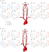
MRI-informed CFD flow and pressure waveforms at selected locations above the carotid bifurcation and in the CoW over a full cardiac cycle. MRI-informed CFD flow waveforms in the cervical arteries are compared to in-vivo PC-MRI flow data. (A) Pre-operative model of patient 1 (B) Pre-operative model of patient 2. RMCA, right middle cerebral artery; RACA, right anterior cerebral artery; RICA, right internal carotid artery; RVA, right vertebral artery; LACA, left anterior cerebral artery; LMCA, left middle cerebral artery; LVA, left vertebral artery; LICA, left internal carotid artery; MRI, magnetic resonance imaging; CFD, computational fluid dynamics; PC-MRI, phase-contrast MRI; CoW, circle of Willis.
Figure 3
Figure 3
Analysis of blood supply in the CoW with Lagrangian particle tracking. Particles are color-coded based on the originating cervical artery. Histograms show the particle count over a full cardiac cycle at each outlet. The accuracy of the MRI-informed CFD models was demonstrated by validating the supply of the intracranial arteries obtained from LPT against territorial perfusion from vessel-selective ASL, which resulted in an overall good qualitative match. A more comprehensive and quantitative validation of the patient models was previously presented (19). LACA, left anterior cerebral artery; RACA, right anterior cerebral artery; LMCA, left middle cerebral artery; RMCA, right middle cerebral artery; LPCA, left posterior cerebral artery; RPCA, right posterior cerebral artery; RICA, right internal carotid artery; LICA, left internal carotid artery; RVA, right vertebral artery; LVA, left vertebral artery; Cow, circle of Willis; MRI, magnetic resonance imaging; CFD, computational fluid dynamics; LPT, Lagrangian particle tracking; ASL, arterial spin labeling.
Figure 4
Figure 4
Comparison of the preoperative and postoperative blood supply in the CoW of patient 2 using Lagrangian particle tracking. Particles are color-coded based on the originating cervical artery. Histograms show the particle count over a full cardiac cycle at the right and left ACA. A close-up shows the flow in the anterior communication artery. LACA, left anterior cerebral artery; RACA, right anterior cerebral artery; RICA, right internal carotid artery; LICA, left internal carotid artery; LVA, left vertebral artery; Cow, circle of Willis; ACA, anterior cerebral artery.
Figure 5
Figure 5
Comparison of preoperative and postoperative mean flow rates in the main arteries of patient 2. Mean flow rates were normalized by the CO. CO, cardiac output; LICA, left internal carotid artery; RICA, right internal carotid artery; LVA, left vertebral artery; RVA, right vertebral artery; LACA, left anterior cerebral artery; RACA, right anterior cerebral artery; LMCA, left middle cerebral artery; RMCA, right middle cerebral artery; LPCA, left posterior cerebral artery; RPCA, right posterior cerebral artery; LSCA, left superior cerebral artery; RSCA, right superior cerebral artery.
Figure 6
Figure 6
Evaluation of the mean pressure gradient ΔP¯ over the stenosis in patients 1 and 2. The pressure distribution in the vasculature is shown at peak systole. ΔP¯ was calculated as the difference between the mean pressures 2 cm proximal and distal to the peak stenosis. RICA, right internal carotid artery; LICA, left internal carotid artery.

References

    1. Flaherty ML, Kissela B, Khoury JC, Alwell K, Moomaw CJ, Woo D, Khatri P, Ferioli S, Adeoye O, Broderick JP, Kleindorfer D. Carotid artery stenosis as a cause of stroke. Neuroepidemiology 2013;40:36-41. 10.1159/000341410 - DOI - PMC - PubMed
    1. Lalla R, Raghavan P, Chaturvedi S. Trends and controversies in carotid artery stenosis treatment. F1000Res 2020. doi:.10.12688/f1000research.25922.1 - DOI - PMC - PubMed
    1. Liebeskind DS. Collateral circulation. Stroke 2003;34:2279-84. 10.1161/01.STR.0000086465.41263.06 - DOI - PubMed
    1. Saba L, Yuan C, Hatsukami TS, Balu N, Qiao Y, DeMarco JK, Saam T, Moody AR, Li D, Matouk CC, Johnson MH, Jäger HR, Mossa-Basha M, Kooi ME, Fan Z, Saloner D, Wintermark M, Mikulis DJ, Wasserman BA; . Carotid Artery Wall Imaging: Perspective and Guidelines from the ASNR Vessel Wall Imaging Study Group and Expert Consensus Recommendations of the American Society of Neuroradiology. AJNR Am J Neuroradiol 2018;39:E9-E31. 10.3174/ajnr.A5488 - DOI - PMC - PubMed
    1. Gupta A, Chazen JL, Hartman M, Delgado D, Anumula N, Shao H, Mazumdar M, Segal AZ, Kamel H, Leifer D, Sanelli PC. Cerebrovascular reserve and stroke risk in patients with carotid stenosis or occlusion: a systematic review and meta-analysis. Stroke 2012;43:2884-91. 10.1161/STROKEAHA.112.663716 - DOI - PMC - PubMed