

The First Case of Fetus in Fetu in Nicaragua: The Management Experience of the Pediatric Neurosurgery Team
- PMID: 36819441
- PMCID: PMC9931383
- DOI: 10.7759/cureus.33835
The First Case of Fetus in Fetu in Nicaragua: The Management Experience of the Pediatric Neurosurgery Team
Abstract
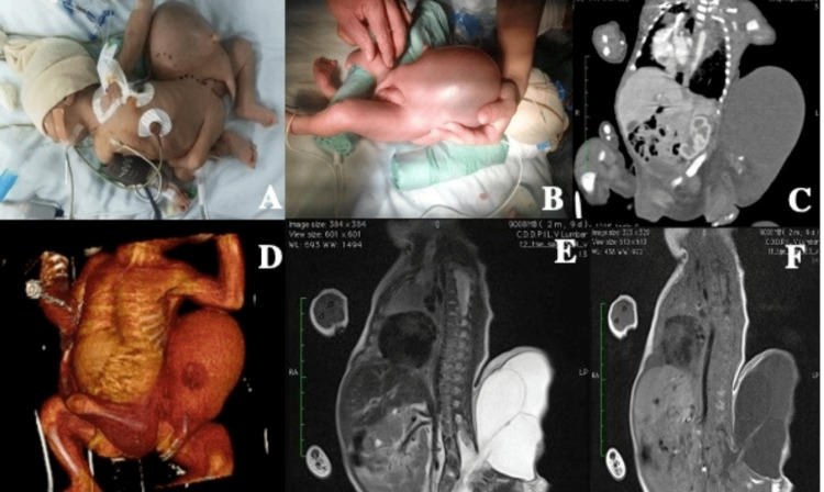
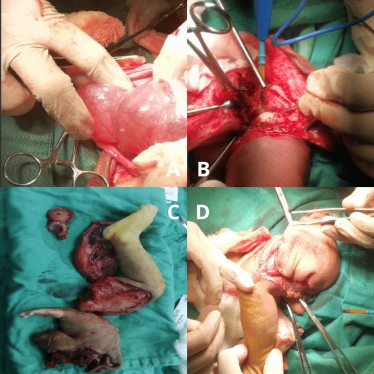
Fetus in fetu (FIF) is a rare congenital anomaly of asymmetric monozygotic twins, where the parasitic twin develops abnormally inside the body of the host twin. In most cases, it is incorporated into the sibling's abdomen, which frequently presents as a retroperitoneal mass. Currently, at least 200 cases have been reported worldwide, being this the first case in Nicaragua. We describe a case of a male newborn, born via cesarean section, with a history of multiple congenital malformations observed via ultrasound examination. At birth, a mass is observed on its dorsum that impresses a skull, but without the presence of bones, with three limbs, two upper and one lower, with an outline located transversely on the pelvic girdle and the presence of two male genitalia with agenesis of the testicles and an accessory kidney. A preoperative diagnosis of FIF and spinal dysraphism was made by computed tomography (CT) and magnetic resonance imaging (MRI). They shared a spinal cord and had the presence of an open spinal defect type meningocele with aberrant roots. After the diagnosis and discussion, the multidisciplinary team proceeded to surgery to perform the separation of the twin (FIF). The subsequent anatomopathological examination revealed that the fetus was anencephalic and had reliable FIF characteristics. The resection was performed followed by the closure of the 430 mL meningocele and complete separation of the spine and the parasitic twin. We present the first case of fetus in fetu in Nicaragua.
Keywords: fetus in fetu; meningocele; neural tube defect; parasitic twin; spina bifida.
Copyright © 2023, Cantarero et al.
Conflict of interest statement
The authors have declared that no competing interests exist.
Figures
Similar articles
-
Ultrasound diagnosis of a retroperitoneal fetus in fetu: A case report.Exp Ther Med. 2023 Apr 27;25(6):284. doi: 10.3892/etm.2023.11983. eCollection 2023 Jun. Exp Ther Med. 2023. PMID: 37206542 Free PMC article.
-
Fetus in Fetu: A Rare Cause of an Abdominal Lump.Cureus. 2024 Oct 14;16(10):e71464. doi: 10.7759/cureus.71464. eCollection 2024 Oct. Cureus. 2024. PMID: 39544589 Free PMC article.
-
Fetus in Fetu: Case Report and Brief Review of Literature on Embryologic Origin, Clinical Presentation, Imaging and Differential Diagnosis.Pol J Radiol. 2017 Jan 30;82:46-49. doi: 10.12659/PJR.899956. eCollection 2017. Pol J Radiol. 2017. PMID: 28217238 Free PMC article.
-
Diagnostic dilemma in a neglected case of fetus-in-fetu solved with Magnetic Resonance Imaging and MDCT--a case report and review of literature.J Radiol Case Rep. 2011;5(10):29-37. doi: 10.3941/jrcr.v5i10.833. Epub 2011 Oct 1. J Radiol Case Rep. 2011. PMID: 22470767 Free PMC article. Review.
-
Fetus in fetu: two case reports and literature review.BMC Pediatr. 2014 Apr 2;14:88. doi: 10.1186/1471-2431-14-88. BMC Pediatr. 2014. PMID: 24693883 Free PMC article. Review.
Cited by
-
Shamrock fetus in fetu: An anesthetic enigma.Med J Armed Forces India. 2025 Mar-Apr;81(2):222-225. doi: 10.1016/j.mjafi.2024.07.009. Epub 2024 Sep 19. Med J Armed Forces India. 2025. PMID: 40496226
-
Fetus In Fetu With Myelomeningocele.Glob Pediatr Health. 2023 Nov 21;10:2333794X231210621. doi: 10.1177/2333794X231210621. eCollection 2023. Glob Pediatr Health. 2023. PMID: 38024466 Free PMC article.
References
-
- Prenatal and postnatal MRI imaging findings of intracranial parasitic fetus: a case report. Zhu K. Childs Nerv Syst. 2021;37:1803–1806. - PubMed
-
- Fetus in fetu--diagnostic criteria and differential diagnosis--a case report and literature review. Brand A, Alves MC, Saraiva C, et al. J Pediatr Surg. 2004;39:616–618. - PubMed
-
- Fetus in fetu or giant epignathus protruding from the mouth. Senyüz OF, Rizalar R, Celayir S, Oz F. J Pediatr Surg. 1992;27:1493–1495. - PubMed
-
- Fetus in fetu. Botell ML, Bermúdez MR. http://scielo.sld.cu/pdf/gin/v39n1/gin08113.pdf Rev Cubana Obstet Ginecol. 2013;39:63–68.
-
- Fetus-in-fetu: a case report and review of the literature. Thakral CL, Maji DC, Sajwani MJ. J Pediatr Surg. 1998;33:1432–1434. - PubMed
Publication types
LinkOut - more resources
Full Text Sources