Review
. 2023 Feb 3:9:1063632.
doi: 10.3389/fmolb.2022.1063632. eCollection 2022.

Hsp70.1 carbonylation induces lysosomal cell death for lifestyle-related diseases

Affiliations
Review

Hsp70.1 carbonylation induces lysosomal cell death for lifestyle-related diseases

Tetsumori Yamashima et al. Front Mol Biosci. .

Abstract

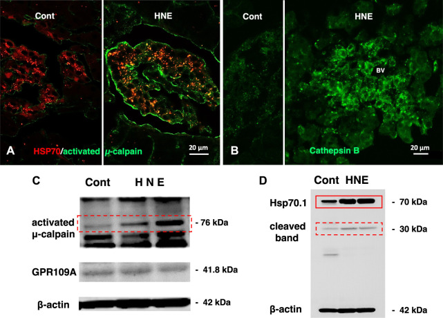
Alzheimer's disease, type 2 diabetes, and non-alcoholic steatohepatitis (NASH) constitute increasingly prevalent disorders. Individuals with type 2 diabetes are well-known to be susceptible to Alzheimer's disease. Although the pathogenesis of each disorder is multifactorial and the causal relation remains poorly understood, reactive oxygen species (ROS)-induced lipid and protein oxidation conceivably plays a common role. Lipid peroxidation product was recently reported to be a key factor also for non-alcoholic steatohepatitis, because of inducing hepatocyte degeneration/death. Here, we focus on implication of the representative lipid-peroxidation product 'hydroxynonenal' for the cell degeneration/death of brain, pancreas, and liver. Since Hsp70.1 has dual roles as a chaperone and lysosomal membrane stabilizer, hydroxynonenal-mediated oxidative injury (carbonylation) of Hsp70.1 was highlighted. After intake of high-fat diets, oxidation of free fatty acids in mitochondria generates ROS which enhance oxidation of ω-6 polyunsaturated fatty acids (PUFA) involved within biomembranes and generate hydroxynonenal. In addition, hydroxynonenal is generated during cooking deep-fried foods with vegetable oils especially containing linoleic acids. These intrinsic and exogenous hydroxynonenal synergically causes an increase in its serum and organ levels to induce Hsp70.1 oxidation. As it is amphiphilic; being water-soluble but displays strong lipophilic characteristics, hydroxynonenal can diffuse within the cells and react with targets like senile and/or atheromatous plaques outside the cells. Hydroxynonenal can deepen and expand lysosomal injuries by facilitating 'calpain-mediated cleavage of the carbonylated Hsp70.1'. Despite the unique anatomical, physiological, and biochemical characteristics of each organ for its specific disease, there should be a common cascade of the cell degeneration/death which is caused by hydroxynonenal. This review aims to implicate hydroxynonenal-mediated Hsp70.1 carbonylation for lysosomal membrane permeabilization/rupture and the resultant cathepsin leakage for inducing cell degeneration/death. Given the tremendous number of worldwide people suffering various lifestyle-related diseases, it is valuable to consider how ω-6 PUFA-rich vegetable oils is implicated for the organ disorder.

Keywords: Alzheimer’s disease; calpain-cathepsin hypothesis; hydroxynonenal; non-alcoholic steatohepatitis; type 2 diabetes.


Conflict of interest statement

The authors declare that the research was conducted in the absence of any commercial or financial relationships that could be construed as a potential conflict of interest.

Figures

FIGURE 1
FIGURE 1
Increased serum hydroxynonenal (4-HNE) level in the patients with type 2 diabetes (T2DM) Panel (A) The serum 4-HNE level was significantly higher in T2DM patients, compared to the non-diabetic subjects (Control). Panels (B) and (C) The simple linear analysis shows that the serum 4-HNE level was positively correlated with HbA1c (B) and fasting glucose (C). Panel (D) The serum 4-HNE level was closely related to the occurrence of T2DM. Reprinted with permission from Lou et al. (2020).
FIGURE 2
FIGURE 2
Molecular markers and hydroxynonenal (HNE) adducts in the hippocampus of Aldh2−/− mice. Panel (A) Immunoblot analysis of hippocampal homogenates from wildtype or Aldh2−/− mice. Aldh2−/− mice showed increase of Alzheimer’s disease-associated markers (top part) and decrease in synaptic markers (bottom part), compared to the wildtype. Panel (B) Aldh2−/− mice showed a significant increase of HNE adducts, compared to the wildtype. APP, amyloid precursor protein; P-tau, hyperphosphorylated tau protein; Casp, caspase; PSD95, postsynaptic density protein 95; CREB, cyclic AMP response element binding protein. Reprinted with permission from D’Souza et al. (2015).
FIGURE 3
FIGURE 3
High hydroxynoneal levels in Alzheimer’s disease (AD) and mild cognitive impairment (MCI). Panel (A) The plasma hydroxynonenal level (HNE) in the patients with Alzheimer’s disease (AD) and the control subjects. (Cited from Rani et al., 2017). Panel (B) Tissue hydroxynonenal concentrations in the hippocampus/parahippocampal gyrus (HPG), superior and middle temporal gyrus (SMTG), and cerebellum (CER) in the patients of MCI, early Alzheimer’s disease (EAD), and age-matched control subjects. Adapted with permission from Williams et al. (2006).
FIGURE 4
FIGURE 4
Electron microphotograph of the lysosomal rupture being observed in the cortical neuron of human Alzheimer patient. Red arrows are lysosomal membrane rupture/permeabilization, which shows a remarkable contrast to the intact lysosome (circles). Apl: autophagolysosome (Reprinted from Yamashima (2020)).
FIGURE 5
FIGURE 5
Calpain activation, Hsp70.1 cleavage, and cathepsin B leakage in the monkeys after the consecutive hydroxynonenal (HNE) injections. Panel (A) Activated μ-calpain immunoreactivity (green) is negligible before HNE injections (Cont), whereas μ-calpain activation occurred after HNE injections (HNE), being consistent with the Western blotting data (Panel (C), activated μ-calpain). After HNE injections, activated μ-calpain immunoreactivity (green) is colocalized with Hsp70.1 immunoreactivity (red), showing a merged color of yellow (HNE, yellow). Panel (B) Cathepsin B is stained as tiny granules in the control Langerhans islet (Cont), whereas stained as coarse granules with the perigranular immunoreactivity after HNE injections (HNE), which indicates lysosomal membrane rupture/permeabilization. Panel (C) By Western blotting, μ-calpain is activated after HNE injections (dot rectangle), compared to the control (Cont). As this anti-μ-calpain antibody recognizes only activated form of μ-calpain, but not inactivated form, positive bands indicate activation of μ-calpain. Panel (D) In response to HNE injections, not only Hsp70.1 main bands (rectangle) but also cleaved Hsp70.1 bands of 30 kDa (dot rectangle) are increased, compared to the control. Reprinted with permission from Boontem and Yamashima (2021).
FIGURE 6
FIGURE 6
Alda-1 in CDAA mice suppresses liver fibrosis (Panel A and lysosomal disintegrity (Panel B). Panels (A, B) rectangles: CDAA mice show fibrosis on the Sirius red staining and lysosomal permeabilization on the Lamp-2 staining, while Alda-1 treatment (CDAA + Alda-1) disclosed decreased immunoreactivity of not only Sirius red and Lamp-2 but also HNE. Panel (C) Western blotting analyses of liver hydroxynonenal protein adducts in CDAA mice (CDAA) and CDAA mice with Alda-1 treatment (CDAA + Alda-1). Alda-1 treatment discloses decreased adducts. Panel (C) Each bands were quantified and shown as relative fold ratios. Adapted with permission from Seike et al. (2022).
FIGURE 7
FIGURE 7
Hydroxynonenal (HNE) induces liver injury in the Japanese macaque monkeys. Panel (A) Macroscopic findings of livers of the control (Cont) and HNE-treated (HNE) monkeys. Black arrows show nodular discoloration. Panel (B) H-E staining and HNE immunostaining of liver tissue from the control group (Cont) and HNE-treated (HNE) group. HNE immunoreactivity was negligible in the control hepatocytes, but was distinct in the latter hepatocytes. Panel (C) Western blotting analyses of the liver HNE protein adducts in the control (Cont) and HNE-treated (HNE) group. P, protein marker. Panel (D) Bands of panel C were quantified and shown as relative fold ratios Panel (E) ALT showed a significant increase after hydroxynonenal injections (HNE), compared to the control (Pre). Adapted with permission from Seike et al. (2022).
FIGURE 8
FIGURE 8
Hydroxynonenal (HNE) is involved in the progression of disease in human NASH. Panel (A) Semi-quantitative assessment of HNE immunoreactivity in the liver tissue of patients with non-fatty liver disease (NAFLD). The density of HNE immunoreactivity was scored into 3 grades: no staining (Grade 0), weak and uniform staining (Grade 1), and intense spots (rectangle) with uniform staining (Grade 2). Panel (B) Immunofluorescence staining of the liver tissue from patients with non-fatty liver and NASH shows that lysosomal membrane permeabilization/rupture was negligible in the former (yellow rectangle), but occurred remarkably in the latter (red rectangle). Blue, DAPI; green, cathepsin B (CTSB); red, Lamp-2. Panel (C) Relationship between the HNE staining score and double-stained granule sizes for Lamp-2 and cathepsin (B). Panel (D) Electron microphotographs of the non-fatty liver and NASH liver. Lysosomes with distinct limiting membrane structures were observed in the non-fatty liver (white arrowhead). In contrast, lysosomes in the NASH liver showed disintegrity of the lysosomal membrane (yellow arrowheads). Panel (E) Western blotting analysis of μ-calpain in the liver tissues of non-fatty liver and NASH, shows an increased activation of μ-calpain in NASH. P, protein marker. Panel (F) Bands of panel E are quantified and shown as relative fold ratios. Adapted with permission from Seike et al. (2022).
FIGURE 9
FIGURE 9
Upregulation, oxidation, and cleavage of Hsp70.1 after transient brain ischemia. Panel (A) Two-dimensional gel electrophoresis with immunoblot detection of carbonylated protein analysis (2D Oxyblot) of the postischemic hippocampal CA1 tissues after immunoprecipitation with anti-Hsp70.1 antibody, shows upregulation of carbonylated Hsp70.1 on the postischemic days 3 (pink) and 5 (blue), compared to the control (black). The specific oxidation index is significantly high on days 3 and 5. Panel (B) Matrix-assisted laser desorption ionization-time of flight/time of flight (MALDI-TOF/TOF) analysis of the upregulated spots with the Mascot search. Both the peptide sequence of the carbonylated peptide ion (459-FELSGIPPAPR*G-470) and the presence of y2 fragment ion atm/z 113.12, indicates that carbonylation occurred at Arg469 in Hsp70.1. R*: Carbonylated arginine Panel (C) In response to hydroxynonenal being generated by ROS, carbonylation occurred at the key site, Arg469 of Hsp70.1. A decrease of its molecular weight from 157.20 to 113.12 is compatible with the insult of carbonylation (Panels A,B,C: cited from Oikawa et al., 2009). Panel (D) In-vitro cleavage of Hsp70.1 by activated μ-calpain in brain tissues from the non-ischemic monkey. It is likely that hydroxynonenal-induced carbonylation facilitates calpain-mediated cleavage of the carbonylated Hsp70.1. Reprinted with permission from Sahara and Yamashim (2010); Yamashima et al. (2014).
FIGURE 10
FIGURE 10
The calpain-cathepsin cascade explaining the molecular mechanism from ω-6 fatty acid-rich PUFA to cell death in lifestyle-related diseases. Diverse G protein-coupled receptors as GPR40/109A/120 in the brain/pancreas/liver are related to Ca2+ mobilization in response to fatty acids. Simultaneously, circumferential oxidative stress and/or deep frying may cause oxidation of ω-6 fatty acid with the resultant generation of hydroxynonenal. Hsp70.1 is a stress-induced protein or lysosomal stabilizer that confer cell protection against diverse stimuli, but its dysfunction caused by calpain-mediated cleavage of carbonylated Hsp70.1 induces diverse cell degeneration via lysosomal rupture and autophagy failure. It is probable that the same disorder may occur for the other lysosomal membrane proteins like Lamp-2. Adapted with permission from Yamashima et al. (2020).

References

    1. Ando Y., Brannstrom T., Uchida K., Nyhlin N., Nasman B., Suhr O., et al. (1998). Histochemical detection of 4-hydroxynonenal protein in Alzheimer amyloid. J. Neurol. Sci. 156, 172–176. 10.1016/s0022-510x(98)00042-2 - DOI - PubMed
    1. Antunes F., Cadenas E., Brunk U. T. (2001). Apoptosis induced by exposure to a low steady-state concentration of H2O2 is a consequence of lysosomal rupture. Biochem. J. 356 (2), 549–555. 10.1042/0264-6021:3560549 - DOI - PMC - PubMed
    1. Arnandis T., Ferrer-Vicens I., García-Trevijano E. R., Miralles V. J., García C., Torres L., et al. (2012). Calpains mediate epithelial-cell death during mammary gland involution: Mitochondria and lysosomal destabilization. Cell. Death Differ. 19, 1536–1548. 10.1038/cdd.2012.46 - DOI - PMC - PubMed
    1. Arnold S. E., Arvanitakis Z., Macauley-Rambach S. L., Koenig A. M., Wang H. Y., Ahima R. S., et al. (2018). Brain insulin resistance in type 2 diabetes and Alzheimer disease: concepts and conundrums. Nat. Rev. Neurol. 14, 168–181. 10.1038/nrneurol.2017.185 - DOI - PMC - PubMed
    1. Attems J., Jellinger K. A. (2014). The overlap between vascular disease and Alzheimer’s disease – lessons from pathology. BMC Med. 12, 206. 10.1186/s12916-014-0206-2 - DOI - PMC - PubMed