

A machine learning tool to improve prediction of mediastinal lymph node metastases in non-small cell lung cancer using routinely obtainable [18F]FDG-PET/CT parameters
- PMID: 36820890
- PMCID: PMC10199849
- DOI: 10.1007/s00259-023-06145-z
A machine learning tool to improve prediction of mediastinal lymph node metastases in non-small cell lung cancer using routinely obtainable [18F]FDG-PET/CT parameters
Abstract
Background: In patients with non-small cell lung cancer (NSCLC), accuracy of [18F]FDG-PET/CT for pretherapeutic lymph node (LN) staging is limited by false positive findings. Our aim was to evaluate machine learning with routinely obtainable variables to improve accuracy over standard visual image assessment.
Methods: Monocentric retrospective analysis of pretherapeutic [18F]FDG-PET/CT in 491 consecutive patients with NSCLC using an analog PET/CT scanner (training + test cohort, n = 385) or digital scanner (validation, n = 106). Forty clinical variables, tumor characteristics, and image variables (e.g., primary tumor and LN SUVmax and size) were collected. Different combinations of machine learning methods for feature selection and classification of N0/1 vs. N2/3 disease were compared. Ten-fold nested cross-validation was used to derive the mean area under the ROC curve of the ten test folds ("test AUC") and AUC in the validation cohort. Reference standard was the final N stage from interdisciplinary consensus (histological results for N2/3 LNs in 96%).
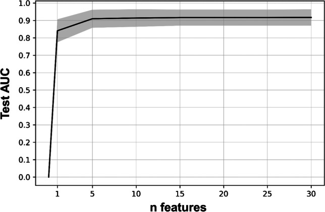
Results: N2/3 disease was present in 190 patients (39%; training + test, 37%; validation, 46%; p = 0.09). A gradient boosting classifier (GBM) with 10 features was selected as the final model based on test AUC of 0.91 (95% confidence interval, 0.87-0.94). Validation AUC was 0.94 (0.89-0.98). At a target sensitivity of approx. 90%, test/validation accuracy of the GBM was 0.78/0.87. This was significantly higher than the accuracy based on "mediastinal LN uptake > mediastinum" (0.7/0.75; each p < 0.05) or combined PET/CT criteria (PET positive and/or LN short axis diameter > 10 mm; 0.68/0.75; each p < 0.001). Harmonization of PET images between the two scanners affected SUVmax and visual assessment of the LNs but did not diminish the AUC of the GBM.
Conclusions: A machine learning model based on routinely available variables from [18F]FDG-PET/CT improved accuracy in mediastinal LN staging compared to established visual assessment criteria. A web application implementing this model was made available.
Keywords: Artificial intelligence; FDG-PET/CT; Lung cancer; Lymph node staging; Machine learning; NSCLC.
© 2023. The Author(s).
Conflict of interest statement
Julian M.M. Rogasch is a participant in the BIH-Charité Digital Clinician Scientist Program funded by the Charité – Universitätsmedizin Berlin, the Berlin Institute of Health, and the German Research Foundation (DFG). Tobias Penzkofer was supported by Berlin Institute of Health (Clinician Scientist Grant, Platform Grant), Ministry of Education and Research (BMBF, 01KX2021, 68GX21001A), German Research Foundation (DFG, SFB 1340/2), Horizon 2020 (952172) and reports research agreements (no personal payments, outside of submitted work) with AGO, Aprea AB, ARCAGY-GINECO, Astellas Pharma Global Inc. (APGD), Astra Zeneca, Clovis Oncology, Inc., Dohme Corp, Holaira, Incyte Corporation, Karyopharm, Lion Biotechnologies, Inc., MedImmune, Merck Sharp, Millennium Pharmaceuticals, Inc., Morphotec Inc., NovoCure Ltd., PharmaMar S.A. and PharmaMar USA, Inc., Roche, Siemens Healthineers, and TESARO Inc., and fees for a book translation (Elsevier). All other authors have no relevant financial or non-financial interests to disclose.
Figures
Comment on
-
Neural networks for nodal staging of non-small cell lung cancer with FDG PET and CT: importance of combining uptake values and sizes of nodes and primary tumor.Radiology. 2014 Jan;270(1):91-8. doi: 10.1148/radiol.13122427. Epub 2013 Oct 28. Radiology. 2014. PMID: 24056403 Free PMC article.
References
-
- Postmus PE, Kerr KM, Oudkerk M, Senan S, Waller DA, Vansteenkiste J, et al. Early and locally advanced non-small-cell lung cancer (NSCLC): ESMO Clinical Practice Guidelines for diagnosis, treatment and follow-up. Ann Oncol. 2017;28:iv1-iv21. 10.1093/annonc/mdx222. - PubMed
Publication types
MeSH terms
Substances
LinkOut - more resources
Full Text Sources
Medical
