Review
. 2023 Jul;43(5):921-932.
doi: 10.1007/s10875-023-01451-5. Epub 2023 Feb 23.

Anti-GM-CSF Neutralizing Autoantibodies in Colombian Patients with Disseminated Cryptococcosis

Affiliations
Review

Anti-GM-CSF Neutralizing Autoantibodies in Colombian Patients with Disseminated Cryptococcosis

Carlos A Arango-Franco et al. J Clin Immunol. 2023 Jul.

Abstract

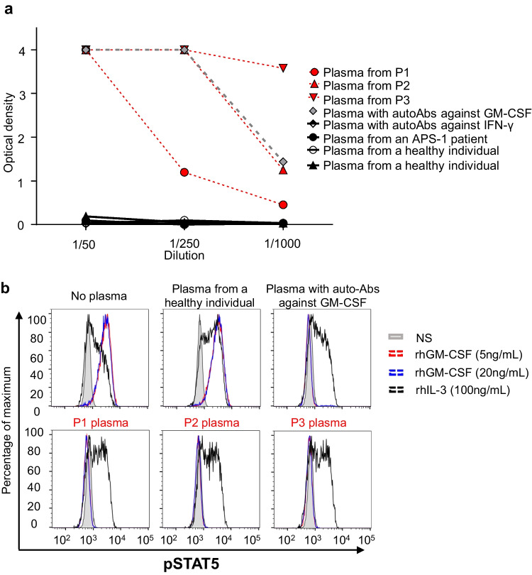
Background: Cryptococcosis is a potentially life-threatening fungal disease caused by encapsulated yeasts of the genus Cryptococcus, mostly C. neoformans or C. gattii. Cryptococcal meningitis is the most frequent clinical manifestation in humans. Neutralizing autoantibodies (auto-Abs) against granulocyte-macrophage colony-stimulating factor (GM-CSF) have recently been discovered in otherwise healthy adult patients with cryptococcal meningitis, mostly caused by C. gattii. We hypothesized that three Colombian patients with cryptococcal meningitis caused by C. neoformans in two of them would carry high plasma levels of neutralizing auto-Abs against GM-CSF.

Methods: We reviewed medical and laboratory records, performed immunological evaluations, and tested for anti-cytokine auto-Abs three previously healthy HIV-negative adults with disseminated cryptococcosis.

Results: Peripheral blood leukocyte subset levels and serum immunoglobulin concentrations were within the normal ranges. We detected high levels of neutralizing auto-Abs against GM-CSF in the plasma of all three patients.

Conclusions: We report three Colombian patients with disseminated cryptococcosis associated with neutralizing auto-Abs against GM-CSF. Further studies should evaluate the genetic contribution to anti-GM-CSF autoantibody production and the role of the GM-CSF signaling pathway in the immune response to Cryptococcus spp.

Keywords: Cryptococcus gattii; Cryptococcus neoformans; Granulocyte–macrophage colony-stimulating factor; Meningitis; Pulmonary alveolar proteinosis (PAP).


Conflict of interest statement

The authors declare that they have no competing interests.

Figures

Fig. 1
Fig. 1
Clinical, microbiologic, and radiologic findings and tissue staining results for patients. Patient 1: a Papular skin lesions on the nasal ala (arrows); b Brain CE-MRI showing multiple nodular lesions affecting the supratentorial areas and the basal ganglia (arrows); c Chest CE-CT scan showing a mediastinal mass surrounding vessels (arrow) and a pulmonary nodule in the upper part of the left lower lobe (arrow); the fistula is not shown; d India ink staining of the mediastinal mass biopsy specimen, showing unstained thick mucopolysaccharide capsules (arrow) from Cryptococcus (40X). Patient 2: e Whole-body CE-MRI: coronal T2-weighted sequence showing hyperintense right supraclavicular fluid collection in the right shoulder affecting the acromioclavicular joint, with infiltration of the bursa and bone (arrows); f Sagittal T2-weighted sequence showing fluid collection extending to the retroperitoneal, dural, and posterior mediastinum, together with spinal cord compression and instability and bone destruction between T9 and T11, with 90% collapse of the space between T10 and T11 (arrows); g Brain CE-MRI showing acute hydrocephalus and basal meningitis with cranial nerve involvement (arrow); h Chest CT-scan showing a mass protruding into the right mediastinal space and bilateral mild pleural effusion (arrow). Patient 3. i Brain MRI sagittal T2-plane showing pathological meningeal enhancement and vasogenic edema (arrow); j Chest CE-CT scan showing an irregular pulmonary nodule in the upper right lobe and a diffuse ground-glass pattern (arrow); k Chest CE-CT scan showing patchy bilateral ground-glass opacities with thickening of the interlobular septa with a “crazy-paving” pattern, predominantly in the lower zone (arrow); l Abundant extracellular proteinaceous periodic acid-Schiff (PAS)-positive material on lung biopsy (100 ×)
Fig. 2
Fig. 2
Anti-GM-CSF neutralizing auto-Abs. a Anti-GM-CSF auto-Ab titers in plasma from the three patients diluted 1:50, 1:250, and 1:1,000 (red), healthy individuals (black) and two patients previously shown to have anti-GM-CSF (grey) or anti-IFN-γ (black and white) auto-Abs, and an APS-1 patient with auto-Abs against IL-17A, IL-17F, IL-22 IFN-α, and IFN-ω (black). b STAT5 phosphorylation (p-STAT5), assessed by flow cytometry, upon the stimulation with GM-CSF or IL-3 of control PBMCs, in the absence of plasma, or in the presence of a 1:10 dilution of plasma from a healthy individual, or from the patients. NS non-stimulated

References

    1. Li J, Vinh DC, Casanova JL, Puel A. Inborn errors of immunity underlying fungal diseases in otherwise healthy individuals. Curr Opin Microbiol. 2017;40:46–57. doi: 10.1016/j.mib.2017.10.016. - DOI - PMC - PubMed
    1. Benedict K, Richardson M, Vallabhaneni S, Jackson BR, Chiller T. Emerging issues, challenges, and changing epidemiology of fungal disease outbreaks. Lancet Infect Dis. 2017;17(12):e403–e411. doi: 10.1016/S1473-3099(17)30443-7. - DOI - PMC - PubMed
    1. Brown GD, Denning DW, Gow NA, Levitz SM, Netea MG, White TC. Hidden killers: human fungal infections. Sci Transl Med. 2012;4(165):165rv13. doi: 10.1126/scitranslmed.3004404. - DOI - PubMed
    1. Pilmis B, Puel A, Lortholary O, Lanternier F. New clinical phenotypes of fungal infections in special hosts. Clin Microbiol Infect. 2016;22(8):681–687. doi: 10.1016/j.cmi.2016.05.016. - DOI - PubMed
    1. Lionakis MS. Primary immunodeficiencies and invasive fungal infection: when to suspect and how to diagnose and manage. Curr Opin Infect Dis. 2019;32(6):531–537. doi: 10.1097/QCO.0000000000000593. - DOI - PubMed

Publication types

Substances