. 2023 Oct;51(5):1383-1398.
doi: 10.1007/s15010-023-01998-w. Epub 2023 Feb 23.

First detected geographical cluster of BoDV-1 encephalitis from same small village in two children: therapeutic considerations and epidemiological implications

Affiliations

First detected geographical cluster of BoDV-1 encephalitis from same small village in two children: therapeutic considerations and epidemiological implications

Leonie Grosse et al. Infection. 2023 Oct.

Abstract

Background: The Borna disease virus (BoDV-1) is an emerging zoonotic virus causing severe and mostly fatal encephalitis in humans.

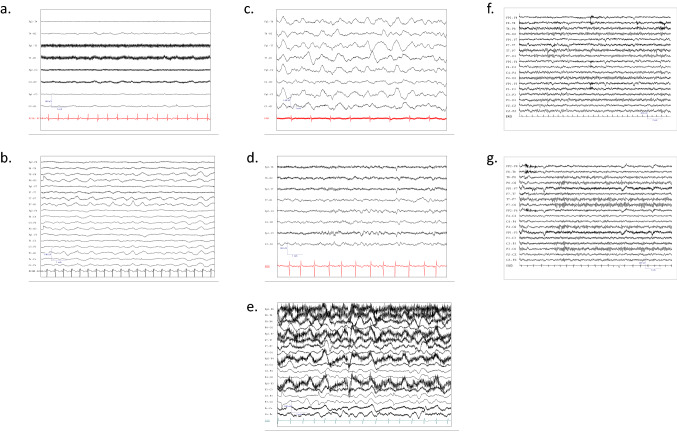
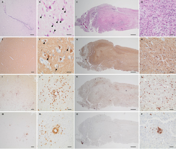
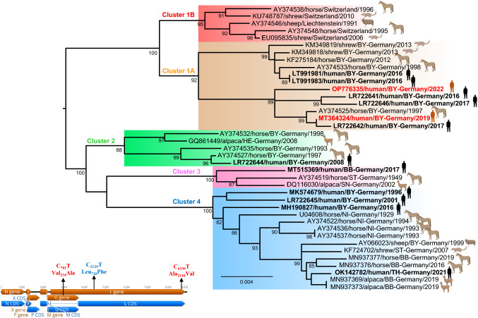
Methods and results: A local cluster of fatal BoDV-1 encephalitis cases was detected in the same village three years apart affecting two children. While the first case was diagnosed late in the course of disease, a very early diagnosis and treatment attempt facilitated by heightened awareness was achieved in the second case. Therapy started as early as day 12 of disease. Antiviral therapy encompassed favipiravir and ribavirin, and, after bioinformatic modelling, also remdesivir. As the disease is immunopathogenetically mediated, an intensified anti-inflammatory therapy was administered. Following initial impressive clinical improvement, the course was also fatal, although clearly prolonged. Viral RNA was detected by qPCR in tear fluid and saliva, constituting a possible transmission risk for health care professionals. Highest viral loads were found post mortem in the olfactory nerve and the limbic system, possibly reflecting the portal of entry for BoDV-1. Whole exome sequencing in both patients yielded no hint for underlying immunodeficiency. Full virus genomes belonging to the same cluster were obtained in both cases by next-generation sequencing. Sequences were not identical, indicating viral diversity in natural reservoirs. Specific transmission events or a common source of infection were not found by structured interviews. Patients lived 750m apart from each other and on the fringe of the settlement, a recently shown relevant risk factor.

Conclusion: Our report highlights the urgent necessity of effective treatment strategies, heightened awareness and early diagnosis. Gaps of knowledge regarding risk factors, transmission events, and tailored prevention methods become apparent. Whether this case cluster reflects endemicity or a geographical hot spot needs further investigation.

Keywords: Borna disease virus; Bornavirus; Epidemiology; Immunosuppression; Transmission; Treatment.


Conflict of interest statement

No conflict of interest concerning this publication has to be disclosed by the authors.

Figures

Fig. 1
Fig. 1
Electroencephalograms of the two cases during the course of disease and healthy controls. Electroencephalogram recording of Case 1 on day 20 of disease (admission to the LMU intensive care unit) showing a general suppression pattern (wave frequency of 2–3/sec (Hertz)) (a), and last recording demonstrating slowing and deterioration of the suppression pattern (1–2 Hertz) (b). Early recording of Case 2 on day 13 showing continuous slowing (3–4 Hertz) (c), temporal improvement with sleep spindles on day 22 (d), and finally on day 46 with delta coma (1–2 Hertz delta waves) (e). Six-year-old healthy matched control awake electroencephalogram recording with normal 7–8 Hertz activity (f). Eleven-year-old matched control awake electroencephalogram recording with normal 8–9 Hertz activity (g). Bottom line represents electrocardiogram (ECG)
Fig. 2
Fig. 2
Brain magnetic resonance imaging of the two pediatric cases over time. Initial scans of Case 1 on day 8 after symptom onset showing signal hyperintensity in the parietal brain regions (fluid-attenuated inversion recovery (FLAIR) sequence) (a) and in the basal ganglia on the left on day 12 (FLAIR sequence) (b). Later scans of the same case on day 15 demonstrating signs of cytotoxic edema (diffusion weighted imaging) (c), and last computed tomography scan on day 29 with diffuse edema (d). Initial scans of Case 2 on day 12 of disease showing signal alterations in temporal brain regions (FLAIR sequence) (e) and in the basal ganglia (FLAIR sequence) (f). Late scans on day 51 demonstrating brain atrophy and signal hyperintensity in the basal ganglia and temporomesial (FLAIR sequence) (g), as well as in the brain stem (FLAIR sequence) (h)
Fig. 3
Fig. 3
Clinical course and treatment overview of Case 1. The figure shows the clinical development during the 29-day-long course of disease, the time elapse until diagnosis, as well as antiviral, immunosuppressive and antibiotic therapy. GCS, Glasgow Coma Scale. IVIG, intravenous immunoglobulins
Fig. 4
Fig. 4
Histology and immunohistochemical staining of brain from Case 1. On H&E, severe parenchymal destruction with almost complete neuronal loss, prominent reactive astrogliosis with enlarged reactive astrocytes with intranuclear viral inclusions (bornavirus-specific Joest-Degen bodies; arrowheads) and diffuse and perivascular accentuated inflammatory infiltration could be observed (A, B). Immunohistochemical staining for BoDV-1 P antigen demonstrated strong diffuse staining with labeling of intranuclear viral inclusions (D, E arrowheads). While infiltrates of CD3-positive T lymphocytes showed a diffuse as well as perivascular accentuated pattern, CD20-positive B lymphocytes were in total less frequent and primarily arranged in perivascular cuffs (C, F). Scale bars: A, C, D, F: 100 µm; B, E: 10 µm
Fig. 5
Fig. 5
Clinical course and treatment overview of Case 2. The figure demonstrates the prolonged clinical course of 67 days, the time elapse until diagnosis, as well as the intensified antiviral and immunosuppressive therapy, and the initial antibiotic treatment. GCS, Glasgow Coma Scale
Fig. 6
Fig. 6
Histology and immunohistochemical staining of brain and olfactory nerve from Case 2. H&E stained sections of the different brain regions sampled (shown here: cingulate gyrus) demonstrated variably pronounced destructive changes with neuronal loss, reactive astrogliosis and inflammatory infiltration (A, B). Joest-Degen nuclear inclusion bodies (arrowheads) could be detected on higher magnification in neurons and astrocytes (B). On immunohistochemistry, strong diffuse positivity for BoDV-1 P antigen including intranuclear Joest-Degen bodies (arrowheads) could be observed (E, F). While CD3-positive T-lymphocytes showed some perivascular accentuation, but were also rather disseminated (I, J), CD20-positive B lymphocytes were arranged in perivascular cuffs (M, N). Similar changes were encountered in the olfactory bulb and tract (respective opposite panels). Stainings C, D: H&E; G, H: BoDV-1-P; K,I: CD3; O, P: CD20. Scale bars: E, I, M: 200 µm; A, C, G, K, O; 100 µm; D, H, J, L, N, P: 50 µm; B, F: 10 µm
Fig. 7
Fig. 7
Maximum likelihood phylogenetic tree of the BoDV-1 strains from the two pediatric patients, based on partial N and P gene nucleotide sequences (~ 1824 bp), as well as depiction of unique mutations of the genome of both strains. Maximum-likelihood bootstrap replicate scores (> 70%) are shown next to the nodes. Taxon information includes GenBank accession number, host species, country of origin (for Germany—federal states (BY-Bavaria; HE-Hesse; BB-Brandenburg; ST-Saxony-Anhalt; SN-Saxony; NI-Lower Saxony; TH-Thuringia) and year of detection are provided. BoDV-1 sequences obtained from the two patients in this study are shown in red. BoDV-1 sequences from humans are depicted in bold. Color code of the clusters are according to [11, 12]. The scale bar indicates nucleotide substitutions per site. The left lower inset shows unique non-synonymous mutation observed in the BoDV-1 genome from the patient OP776335 (mutations in red) and patient MT364324 (mutation in blue)

Similar articles

Cited by

References

    1. Dürrwald R, Kolodziejek J, Weissenböck H, Nowotny N. The bicolored white-toothed shrew Crocidura leucodon (HERMANN 1780) is an indigenous host of mammalian Borna disease virus. PLoS ONE. 2014;9:e93659. doi: 10.1371/journal.pone.0093659. - DOI - PMC - PubMed
    1. Nobach D, Bourg M, Herzog S, Lange-Herbst H, Encarnação JA, Eickmann M, Herden C. Shedding of infectious Borna disease virus-1 in living bicolored white-toothed shrews. PLoS ONE. 2015;10:e0137018. doi: 10.1371/journal.pone.0137018. - DOI - PMC - PubMed
    1. Weissenböck H, Bagó Z, Kolodziejek J, Hager B, Palmetzhofer G, Dürrwald R, Nowotny N. Infections of horses and shrews with Bornaviruses in Upper Austria: a novel endemic area of Borna disease. Emerg Microbes Infect. 2017;6:e52. doi: 10.1038/emi.2017.36. - DOI - PMC - PubMed
    1. Schlottau K, Forth L, Angstwurm K, Höper D, Zecher D, Liesche F, Hoffmann B, Kegel V, Seehofer D, Platen S, Salzberger B, Liebert UG, Niller HH, Schmidt B, Matiasek K, Riemenschneider MJ, Brochhausen C, Banas B, Renders L, Moog P, Wunderlich S, Seifert CL, Barreiros A, Rahmel A, Weiss J, Tappe D, Herden C, Schmidt-Chanasit J, Schwemmle M, Rubbenstroth D, Schlegel J, Pietsch C, Hoffmann D, Jantsch J, Beer M. Fatal encephalitic Borna disease virus 1 in solid-organ transplant recipients. N Engl J Med. 2018;379:1377–1379. doi: 10.1056/NEJMc1803115. - DOI - PubMed
    1. Korn K, Coras R, Bobinger T, Herzog SM, Lücking H, Stöhr R, Huttner HB, Hartmann A, Ensser A. Fatal encephalitis associated with Borna disease virus 1. N Engl J Med. 2018;379:1375–1377. doi: 10.1056/NEJMc1800724. - DOI - PubMed