. 2023 Apr 10;8(7):e162907.
doi: 10.1172/jci.insight.162907.

Reversible epigenetic alterations mediate PSMA expression heterogeneity in advanced metastatic prostate cancer

Affiliations

Reversible epigenetic alterations mediate PSMA expression heterogeneity in advanced metastatic prostate cancer

Erolcan Sayar et al. JCI Insight. .

Abstract

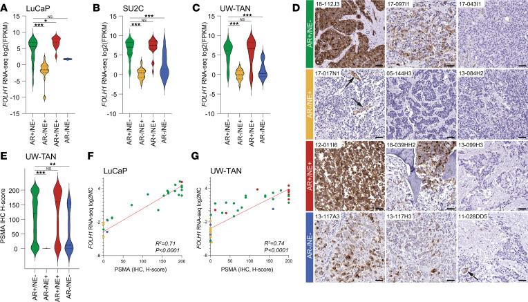
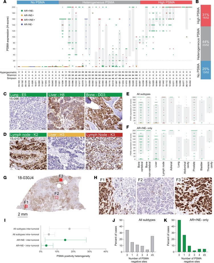
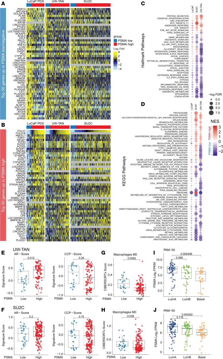
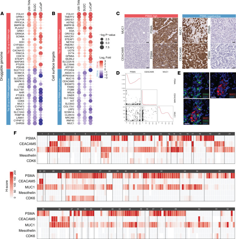
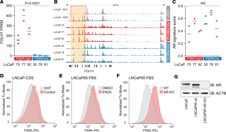
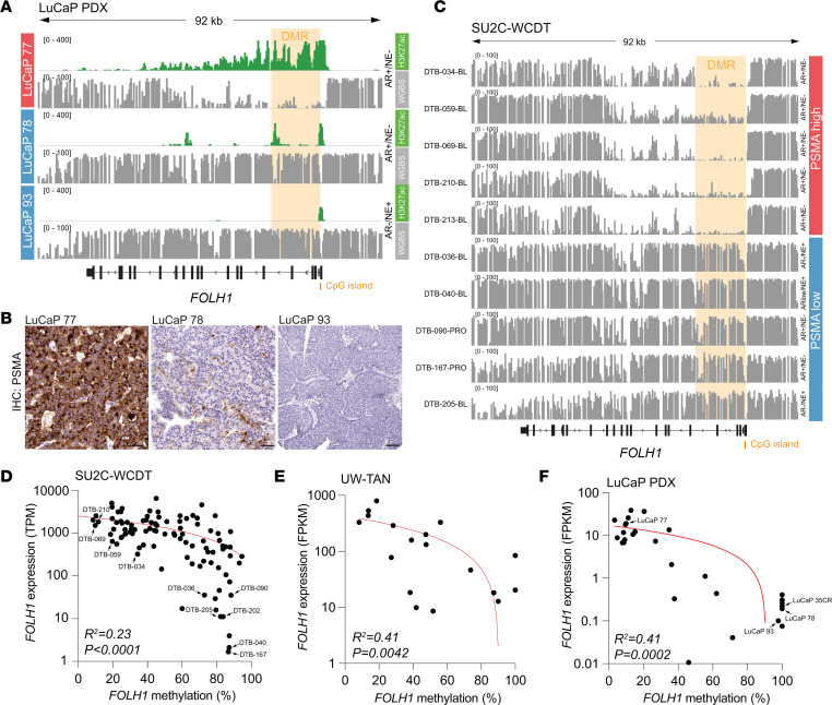
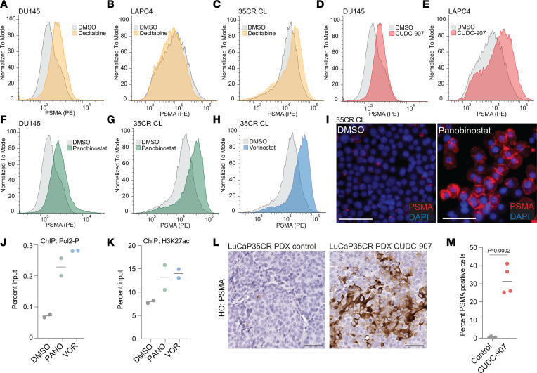
Prostate-specific membrane antigen (PSMA) is an important cell surface target in prostate cancer. There are limited data on the heterogeneity of PSMA tissue expression in metastatic castration-resistant prostate cancer (mCRPC). Furthermore, the mechanisms regulating PSMA expression (encoded by the FOLH1 gene) are not well understood. Here, we demonstrate that PSMA expression is heterogeneous across different metastatic sites and molecular subtypes of mCRPC. In a rapid autopsy cohort in which multiple metastatic sites per patient were sampled, we found that 13 of 52 (25%) cases had no detectable PSMA and 23 of 52 (44%) cases showed heterogeneous PSMA expression across individual metastases, with 33 (63%) cases harboring at least 1 PSMA-negative site. PSMA-negative tumors displayed distinct transcriptional profiles with expression of druggable targets such as MUC1. Loss of PSMA was associated with epigenetic changes of the FOLH1 locus, including gain of CpG methylation and loss of histone 3 lysine 27 (H3K27) acetylation. Treatment with histone deacetylase (HDAC) inhibitors reversed this epigenetic repression and restored PSMA expression in vitro and in vivo. Collectively, these data provide insights into the expression patterns and regulation of PSMA in mCRPC and suggest that epigenetic therapies - in particular, HDAC inhibitors - can be used to augment PSMA levels.

Keywords: Drug therapy; Epigenetics; Oncology; Prostate cancer.


Conflict of interest statement

Conflict of interest: PSN served as a paid advisor for Bristol Myers Squibb, Pfizer, and Janssen. LDT is a cofounder and has equity in Alpenglow biosciences. NHB is an inventor on patents assigned to Cornell University for anti-PSMA antibody technology aA list of all patents can be found at: patentscope.wipo.int, Search term: Neil H. Bander). He is a founder, director, and advisor to Convergent Therapeutics Inc., to which PSMA antibody technology has been licensed. He is also a founder, director, and advisor of XenImmune Therapeutics Inc. EC received sponsored research funding from AbbVie, Astra Zeneca, Foghorn, Kronos, MacroGenics, Bayer Pharmaceuticals, Forma Pharmaceuticals, Janssen Research, Gilead, Arvina, and Zenith Epigenetics. MTS is a paid consultant/received honoria from Sanofi, AstraZeneca, PharmaIN, and Resverlogix and has received research funding from Zenith Epigenetics, Bristol Myers Squibb, Merck, Immunomedics, Janssen, AstraZeneca, Pfizer, Madison Vaccines, Hoffman-La Roche, Tmunity, SignalOne Bio, and Ambrx Inc. JEH reports consultant fees from Seagen and research support from Regeneron and Dendreon paid to her institution. FYF serves on the Scientific Advisory Board of Artera, BlueStar Genomics, and SerImmune and is a paid consultant Astellas, Bayer, Blue Earth Diagnostics, Foundation Medicine, Janssen, Myovant, Novartis, and Roivant.

Figures

Figure 1
Figure 1. PSMA expression patterns differ between molecular subtypes of lethal metastatic prostate cancer.
(AC) Violin plots show distribution of FOLH1 expression (log2 FPKM) across molecular subtypes (AR+/NE [green], AR/NE+ [yellow], AR+/NE+ [red], and AR/NE [blue]), determined by RNA-Seq in LuCaP PDX (n = 126) (A), SU2C (n = 270) (B), and UW-TAN (n = 172) tumors (C) (see Supplemental Table 1 for a breakdown of samples across all molecular phenotypes). (D) Representative micrographs of PSMA IHC in different molecular subtypes. Arrows indicate PSMA-positive endothelial cells in cases with absence of tumor cell–specific PSMA expression. (E) Violin plot depicts PSMA-expression H-scores from the UW-TAN cohort (n = 636). (F and G) Correlation plots show a significant positive association (R2 based on Pearson correlation) between PSMA protein expression (by IHC) and FOLH1 mRNA expression (by RNA-seq) in LuCaP (n = 25) (F) and UW-TAN samples (n = 50) (G). Dot colors indicate molecular phenotypes, as above. Scale bars: 50 μm. *P < 0.05; **P < 0.001; ***P < 0.0001, based on Wilcoxon rank tests.
Figure 2
Figure 2. Inter- and intrapatient PSMA expression heterogeneity.
(A) Dot and box plot show the distribution of PSMA protein expression H-scores in 52 cases from the UW-TAN rapid autopsy cohort (total sample n = 636). Each dot represents a tumor sample; the color codes indicate the molecular subtype (AR+/NE [green], AR/NE+ [yellow], AR+/NE+ [red] and AR/NE [blue]). Gray shadings show interquartile ranges. (B) Summary of frequencies of cases with uniformly low/negative PSMA expression (all sites H-score ≤ 20), heterogeneous PSMA expression (both H-scores ≤ 20 and H-score > 20) and uniformly high PSMA expression (all sites with H-scores > 20). (C) Representative micrographs of PSMA expression patterns in 3 anatomically distinct metastatic sites (all AR+/NE) from 1 case (case 15-096). (D) PSMA expression in a case with divergent subtypes (case 15-010). (E and F) Distribution of PSMA expression H-scores across different organ sites in all tumors (E) and AR+/NE tumors (F). (G) PSMA expression heterogeneity within a metastatic lesion (intratumoral heterogeneity). Left panel shows low power (1×) view of a full-face tumor section. Areas with divergent PSMA expression are indicated (F1–F3). (H) High-power view (20×) of 3 tumor foci (F1–F3 from G) showing high level intratumoral expression heterogeneity. (I) Mean (95% CI) hypergeometric PSMA expression heterogeneity indices across different metastatic sites in a given patient (intertumoral heterogeneity) and within a metastatic site (intratumoral heterogeneity) for the entire cohort (gray) and AR+/NE tumors (green). (J and K) Percentage of cases with no (0), 1, 2, 3, 4 or ≥ 5 metastatic sites with a PSMA H-score ≤ 20 (PSMA-negative metastatic sites). Numbers are shown separately for the entire cohort (gray bars) (J) and in AR+/NE tumors only (green bars) (K). Scale bars: 50 μm.
Figure 3
Figure 3. Tumors with low/negative PSMA expression show distinct expression changes.
(A and B) Heatmap showing the top 50 genes upregulated in AR+/NE tumors from the LuCaP PDX (n = 82), UW-TAN (n = 109), and SU2C (n = 182) cohorts with low/negative PSMA expression (A) and high PSMA expression (B). (C and D) Gene set enrichment analyses using Hallmark Pathways (C) and KEGG Pathways (D) show gene sets enriched in PSMA high (red) and PSMA-low/negative (blue) tumors. NES denotes normalized enrichment score. (E and F) Comparisons of AR signaling activity (AR-score) and cell proliferation (CCP-score) using gene set variation analyses (Supplemental Figures 12 and 13) between PSMA-low/negative and PSMA-high AR+/NE tumors in the UW-TAN (E) and SU2C (F) cohorts. (G and H) CIBERSORTx analyses demonstrate differences in macrophage infiltration between PSMA high and PSMA low/negative tumors in UW-TAN (G) and SU2C (H) cohorts. (I and J) Differences in PSMA expression based on luminal A, luminal B, and basal PAM-50 status in UW-TAN (I) and SU2C (J) cohorts. P values are based Wilcoxon rank tests.
Figure 4
Figure 4. PSMA-low/negative tumors show targetable alterations.
(A) Heatmap of top 20 differentially expressed genes with annotated drug target properties from the druggable genome database (rank ordered based on fold expression difference) between PSMA-high (red) and PSMA-low/negative (blue) AR+/NE tumors in UW-TAN, SU2C, and LuCaP PDX cohorts. (B) Top 20 differentially expressed genes encoding for cell surface proteins between PSMA-high (red) and PSMA-low/negative (blue) tumors in UW-TAN, SU2C, and LuCaP PDX. Heatmaps are sorted by rank order based on mean fold change differences, and directionality is color coded: red, higher in PSMA-high; blue, higher in PSMA-low/negative. (C) Representative micrographs of CEACAM5 and MUC1 IHC in PSMA-negative/low and PSMA-high tumors. (D) Correlation plots for PSMA, MUC1, and CEACAM5 protein expression. (E) Dual fluorescence images showing distinct cell population labeling for MUC1 (red) and PSMA (green). (F) Heatmaps of PSMA, CEACAM5, MUC1, mesothelin, and CDK6 expression based on IHC H-scores across 289 metastatic sites in 52 patients. Expression scores for each protein target are color coded from light gray to red. White boxes indicate missing data. Each colored box represents a metastatic site; black boxes outline each case (see Supplemental Table 8 for UW-TAN case identifiers). Scale bars: 50 μm.
Figure 5
Figure 5. AR-mediated changes in PSMA expression.
(A) FOLH1 mRNA expression in AR+ LuCaP PDX lines. (B) AR ChIP-Seq tracks in LNCaP cell line and LuCaP PDX tissues show AR recruitment at the FOLH1 locus. Red box highlights dihydrotestosterone-induced (DHT-induced) peaks in LNCaP cells. (C) Distribution of 10-gene AR signature (23) across LuCaP models. P values are based on 2-tailed t tests. (D and E) Density plots show PSMA cell surface expression in LnCaP and LNCaP95 (as indicated) in the absence or presence of 10 nM DHT (D) or 10μM enzalutamide (ENZA) (E) for 6 days. (F) PSMA cell surface expression in LNCaP95 AR-KO and parental WT cells. (G) Western blot shows complete loss of AR protein expression in AR-KO cells (40).
Figure 6
Figure 6. Epigenetic changes enforce silencing of FOLH1/PSMA.
(A) Whole-genome bisulfite sequencing (WGBS) tracks from PSMA-high (LuCaP 77, red) and PSMA-negative (LuCaP 78 and 93, blue) tumors reveal a differentially methylated region (DMR) encompassing the first 14 kb of the FOLH1 gene and inverse enrichment for H3K27ac. (B) Micrographs of PSMA IHC of LuCaP 77, LuCaP 78, and LuCaP 93 demonstrate the difference in PSMA expression. (C) Representative WGBS tracks of mCRPC tumors from the SU2C-WCDT cohort show gain of methylation in the DMR in PSMA-low/negative tumors. (DF) Scatter plots show the correlation between FOLH1 expression (based on RNA-Seq) and DMR methylation derived from WGBS (SU2C-WCDT, n = 98) (D) and targeted COMPARE-MS analyses UW-TAN (n = 18) (E) and LuCaP (n = 29) (F) cohorts. Curves were fit by linear regression, and R2 and P values were derived by Pearson correlation.
Figure 7
Figure 7. Pharmacologic epigenetic modifiers reverse PSMA silencing.
(AC) Density plots show PSMA cell surface expression in DU145 (A), LAPC4 (B), and LuCaP 35CR cell line (CL) (C) cells treated for 6 days with vehicle control (DMSO) or 500 nM decitabine (DAC). (DH) PSMA expression in DU145, LAPC4, and LuCaP 35CR CL cells treated for 6 days with panobinostat (PANO, 10 nM), CUDC-907 (50 nM), or vorinostat (VOR, 1 μM). (I) Representative micrographs of cytospin preparations of LuCaP 35CR CL treated with DMSO or panobinostat (10 nM) stained for PSMA (red) and DAPI (blue). (J and K) Chromatin immunoprecipitation studies show serine 5 phosphorylated RNA polymerase 2 (Pol2-P) (J) and H3K27ac (K) enrichment normalized to input in LuCaP 35CR CL treated with vorinostat (VOR, 1 μM), panobinostat (PANO, 10 nM), or DMSO. (L) Micrographs of LuCaP 35CR PDX tumors stained for PSMA grown in mice treated with solvent control (30% captisol) or CUDC-907 at a dose of 75 mg/kg/day for 21 days. (M) Percent of PSMA-positive cells in control and CUDC-907 treated tumors (n = 4 per group). P value are based on 2-tailed t tests. Scale bars: 50 μm.

References

    1. Sheehan B, et al. Prostate-specific membrane antigen biology in lethal prostate cancer and its therapeutic implications. Eur Urol Focus. 2022;8(5):1157–1168. doi: 10.1016/j.euf.2021.06.006. - DOI - PubMed
    1. Nauseef JT, et al. Emerging prostate-specific membrane antigen-based therapeutics: small molecules, antibodies, and beyond. Eur Urol Focus. 2021;7(2):254–257. doi: 10.1016/j.euf.2021.02.006. - DOI - PubMed
    1. Giraudet A-L, et al. PSMA targeting in metastatic castration-resistant prostate cancer: where are we and where are we going? Ther Adv Med Oncol. 2021;13:17588359211053898. doi: 10.1177/17588359211053898. - DOI - PMC - PubMed
    1. Lawhn-Heath C, et al. Prostate-specific membrane antigen PET in prostate cancer. Radiology. 2021;299(2):248–260. doi: 10.1148/radiol.2021202771. - DOI - PubMed
    1. Sartor O, et al. Lutetium-177-PSMA-617 for metastatic castration-resistant prostate cancer. N Engl J Med. 2021;385(12):1091–1103. doi: 10.1056/NEJMoa2107322. - DOI - PMC - PubMed

Publication types

Substances