. 2023 Sep 5;25(9):1605-1616.
doi: 10.1093/neuonc/noad047.

The development of a rapid patient-derived xenograft model to predict chemotherapeutic drug sensitivity/resistance in malignant glial tumors

Affiliations

The development of a rapid patient-derived xenograft model to predict chemotherapeutic drug sensitivity/resistance in malignant glial tumors

Martine Charbonneau et al. Neuro Oncol. .

Abstract

Background: High-grade gliomas (HGG) are aggressive brain tumors associated with short median patient survival and limited response to therapies, driving the need to develop tools to improve patient outcomes. Patient-derived xenograft (PDX) models, such as mouse PDX, have emerged as potential Avatar platforms for personalized oncology approaches, but the difficulty for some human grafts to grow successfully and the long time required for mice to develop tumors preclude their use for HGG.

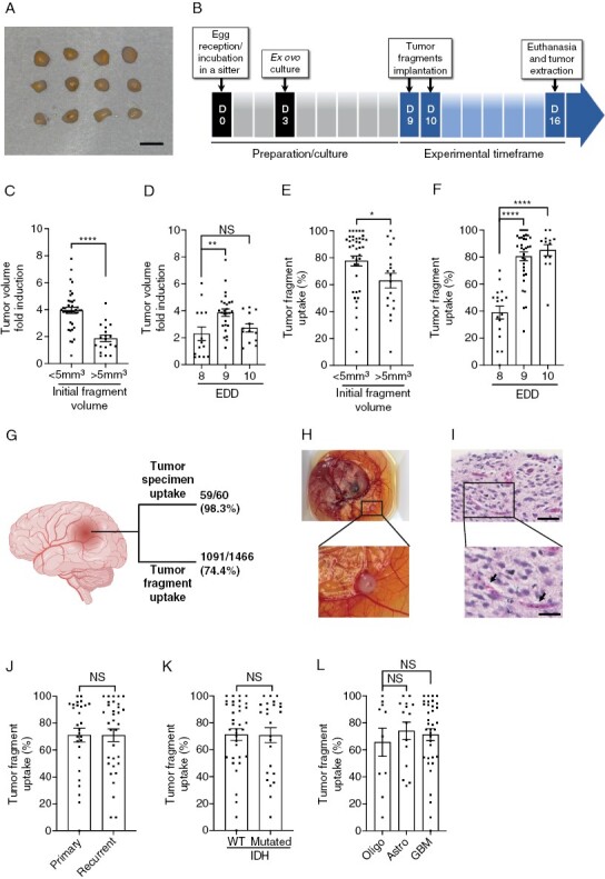
Methods: We used a rapid and efficient ex-ovo chicken embryo chorioallantoic membrane (CAM) culture system to evaluate the efficacy of oncologic drug options for HGG patients.

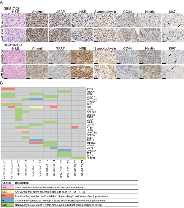
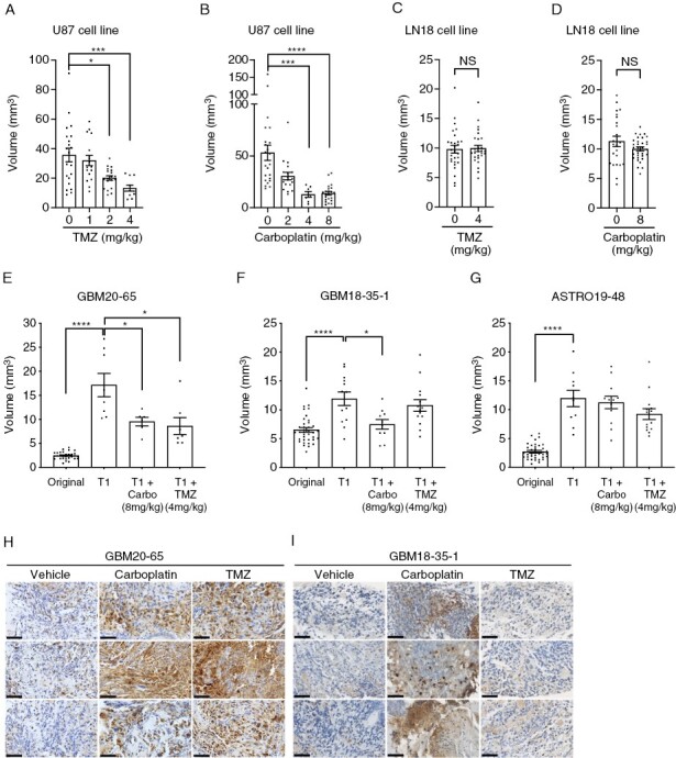
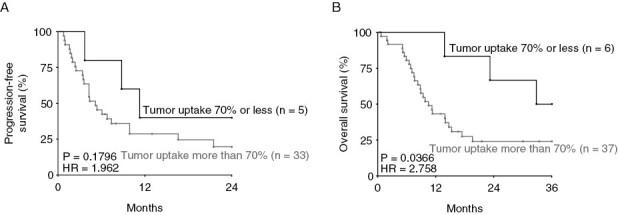
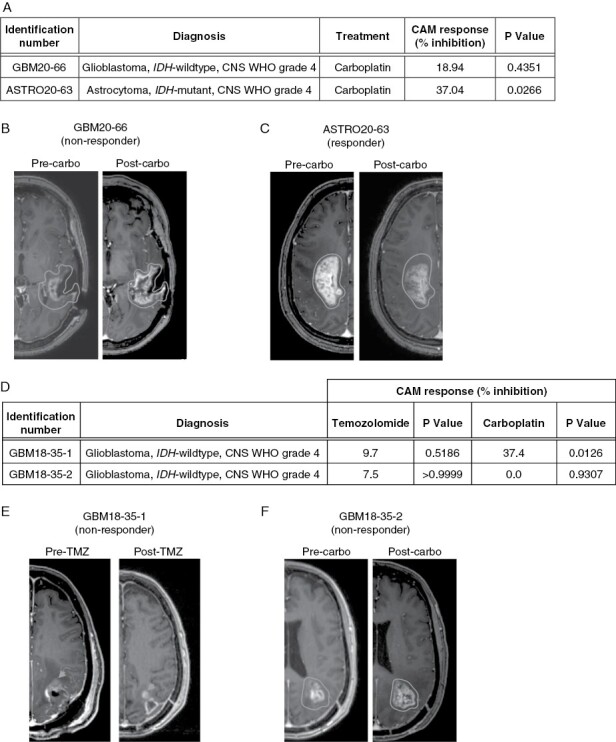
Results: Implantation of fresh glioma tissue fragments from 59 of 60 patients, that include difficult-to-grow IDH-mutated samples, successfully established CAM tumor xenografts within 7 days, with a tumor take rate of 98.3%. These xenografts faithfully recapitulate the histological and molecular characteristics of the primary tumor, and the ability of individual fragments to form tumors was predictive of poor patient prognosis. Treatment of drug-sensitive or drug-resistant xenografts indicates that the CAM-glioma assay enables testing tumor sensitivity to temozolomide and carboplatin at doses consistent with those administered to patients. In a proof-of-concept study involving 14 HGG patients, we observed a correlation of 100% between the CAM xenograft response to temozolomide or carboplatin and the clinical response of patients.

Conclusion: The CAM-glioma model is a fast and reliable assay that has the potential to serve as a complementary model to drug discovery and a real-time Avatar platform to predict the best treatment for HGG patients.

Keywords: Avatar model; chick chorioallantoic membrane assay; glioblastoma; patient-derived xenograft model; personalized medicine.


Figures

Figure 1.
Figure 1.
Optimization of the glioma CAM-PDX model. (A) Representative images of the tumor fragments used for implantation. Scale bar = 2mm. (B) Timeline of the procedure. (C and D) Xenograft volume 6 or 7 days post-implantation based on initial fragment size (C) and embryonic development day (EDD) of implantation (D). (E and F) Tumor uptake as a function of initial fragment size (E) and EDD of implantation (F). (G) Tumor specimen and tumor fragment uptake on CAM. Created with BioRender.com (H) Representative image and (I) representative H&E staining of a CAM-glioma xenograft. Arrows show chicken blood vessels. Scale bar = 50 μm. Zoom in scale bar = 25 μm. (J–L) Tumor take rate according to primary or recurrent tumors (J), IDH status (K), or cell types/grades (L) of gliomas. Oligo: oligodendroglioma, Astro: astrocytoma, GBM: glioblastoma. Values are expressed as mean ± SEM. *P < .05, **P < .01, ****P < .0001, (C,E,J,K) Mann-Whitney test, (D,F,L) Kruskal-Wallis test.
Figure 2.
Figure 2.
Histopathological and genetic characteristics of original and xenografted tumors. (A) Representative images of H&E, vimentin, GFAP, CD44, nestin, synaptophysin, NSE, and Ki67 staining of the original and xenografted tumors; arrows show Ki67 positive cells; N = 5. Scale bar = 50 μm. (B) Mutations in 28 glioblastoma-associated genes identified by whole-exome sequencing of parental (T0) and xenografted (T1) tumors; N = 6. ASTRO: astrocytoma, GBM: glioblastoma, OLIG: oligodendroglioma.
Figure 3.
Figure 3.
Effect of drug treatment on glioma xenografts grown on CAM. (A–D) Tumor volume of xenografts derived from U-87 MG or LN18 cell lines treated with different doses of (A and C) temozolomide (TMZ) (N = 3) or (B and D) carboplatin (Carbo) (N = 3). (E–G) Tumor volume of original glioma fragments (Original) or CAM xenografts (T1) treated with vehicle, carboplatin or TMZ for patient GBM20-65 (E), GBM18-35-1 (F), and ASTRO19-48 (G) and (H,I) representative images of cleaved caspase 3 staining for three separate xenografts per group (n = 3–7). GBM: glioblastoma, ASTRO: astrocytoma. Scale bar = 50 μm. Bars = mean ± SEM. *P < .05, **P < .01, ***P < .001, ****P < .0001, (A and B) Kruskall-Wallis test, (C–G) Mann-Whitney test.
Figure 4.
Figure 4.
Prognostic value of the ability for glioma specimens to form xenografts in the CAM model. Kaplan-Meier analysis of (A) progression-free and (B) OS in glioma patients with high > 70% (n = 33–37) and low < 70% (n = 5–6) tumor fragment uptake on CAM, a cut-off chosen for maximal discrimination between high- and low-grade patients. HR: hazard ratio (logrank), Gehan-Breslow-Wilcoxon test.
Figure 5.
Figure 5.
CAM-glioma and clinical response to chemotherapeutics. (A) Xenograft response to chemotherapeutic drugs for a non-responder (GBM20-66) and a responder (ASTRO20-63) PDX model. (B and C) Associated pre-and post-treatment MRI images for (B) GBM20-66 and (C) ASTRO20-63 patients. (D) Xenograft response to chemotherapeutic agents for two PDX models established from a single patient. (E and F) Pre- and post-treatment MRI images for the patient associated with (E) GBM18-35-1 and (F) GBM18-35-2 samples. Mann-Whitney test.

References

    1. Stupp R, Brada M, van den Bent MJ, Tonn J-C, Pentheroudakis G, ESMO Guidelines working group. high-grade glioma: ESMO Clinical Practice Guidelines for diagnosis, treatment and follow-up. Ann Oncol. 2014;25(suppl 3):iiiiii9393–iii101. - PubMed
    1. Stupp R, Mason WP, van den Bent MJ, et al. ; European Organisation for Research and Treatment of Cancer Brain Tumor and Radiotherapy Groups. Radiotherapy plus concomitant and adjuvant temozolomide for glioblastoma. N Engl J Med. 2005;352(10):987–996. - PubMed
    1. Stupp R, Taillibert S, Kanner AA, et al. . Maintenance therapy with tumor-treating fields plus temozolomide vs temozolomide alone for glioblastoma: a randomized clinical trial. JAMA. 2015;314(23):2535–2543. - PubMed
    1. Patel AP, Tirosh I, Trombetta JJ, et al. . Single-cell RNA-seq highlights intratumoral heterogeneity in primary glioblastoma. Science. 2014;344(6190):1396–1401. - PMC - PubMed
    1. Staedtke V, Dzaye OD, Holdhoff M.. Actionable molecular biomarkers in primary brain tumors. Trends Cancer. 2016;2(7):338–349. - PMC - PubMed

Publication types