. 2023 Mar 17;18(3):518-527.
doi: 10.1021/acschembio.2c00862. Epub 2023 Feb 23.

Rapid Single-Shot Synthesis of the 214 Amino Acid-Long N-Terminal Domain of Pyocin S2

Affiliations

Rapid Single-Shot Synthesis of the 214 Amino Acid-Long N-Terminal Domain of Pyocin S2

Azin Saebi et al. ACS Chem Biol. .

Abstract

The impermeable outer membrane of Pseudomonas aeruginosa is bypassed by antibacterial proteins known as S-type pyocins. Because of their properties, pyocins are investigated as a potential new class of antimicrobials against Pseudomonas infections. Their production and modification, however, remain challenging. To address this limitation, we employed automated fast-flow peptide synthesis for the rapid production of a pyocin S2 import domain. The N-terminal domain sequence (PyS2NTD) was synthesized in under 10 h and purified to yield milligram quantities of the desired product. To our knowledge, the 214 amino acid sequence of PyS2NTD is among the longest peptides produced from a "single-shot" synthesis, i.e., made in a single stepwise route without the use of ligation techniques. Biophysical characterization of the PyS2NTD with circular dichroism was consistent with the literature reports. Fluorescently labeled PyS2NTD binds to P. aeruginosa expressing the cognate ferripyoverdine receptor and is taken up into the periplasm. This selective uptake was validated with confocal and super resolution microscopy, flow cytometry, and fluorescence recovery after photobleaching. These modified, synthetic S-type pyocin domains can be used to probe import mechanisms of P. aeruginosa and leveraged to develop selective antimicrobial agents that bypass the outer membrane.


Figures

Figure 1.
Figure 1.. PyS2NTD was rapidly synthesized in 9.2 h using AFPS.
(A) Schematic representation of the automated flow synthesis and biological function of PyS2NTD. (B) The sequence of PyS2NTD with cysteine (Cys216) added for bioconjugation. (C) The UV absorbance trace at 310 nm from flow synthesis showed the Fmoc-deprotection peaks remained comparable in height and width throughout the synthesis. Coupling steps are also shown and produce a saturated signal, just before each Fmoc-deprotection peak. (D) Analytical RP-HPLC from absorbance at 214 nm and (E) LC-MS mass spectrum of crude PyS2NTD with deconvoluted mass spectrum inset. (F) RP-HPLC of purified PyS2NTD with absorbance at 214 nm and (G) LC-MS mass spectrum of the purified PyS2NTD with deconvoluted mass spectrum inset. For additional information on synthesis, cleavage, and purification conditions see the Methods section.
Figure 2.
Figure 2.. Biophysical characterization of synthetic PyS2NTD after SEC-based folding.
(A) Crystal structure of PyS2NTD (residues 11–205, PDB: 5ODW) displays primarily α-helices and a short antiparallel β-hairpin. (B) Chromatogram of SEC purification of PyS2NTD, showing absorbance at 214 nm, where the fractions containing the protein (shaded in gray) were pooled to yield 0.73 mg of material (84% yield). (C) RP-HPLC of folded PyS2NTD showing absorbance at 214 nm and (D) LC-MS of the folded PyS2NTD with deconvoluted mass spectrum in inset. (E) Far-UV CD spectrum of the purified synthetic PyS2NTD. (F) Thermal melt curve for synthetic PyS2NTD in the temperature range of 25–70 °C. The melting temperature (Tm) was determined to be 49 ± 2 °C (mean ±95% CI), similar to the 50 °C value reported in literature. See Supporting Information and Methods for more on folding, purification, and CD.
Figure 3.
Figure 3.. Synthetic PyS2NTD was conjugated to fluorophore AZDye 488 and folded.
(A) Conjugation and folding scheme of PyS2NTD. (B) Chromatogram of SEC purification of PyS2–488, showing fluorescence signal with excitation at 280 nm and emission at 325 nm, where the fractions containing the protein (shaded in gray) were pooled to yield 234 μg of material corresponding to 24% combined reaction and folding recovery yield. (C) RP-HPLC of folded PyS2–488 conjugate showing absorbance at 214 nm and (D) LC-MS of the folded PyS2–488 conjugate with deconvoluted mass spectrum inset. See Supporting Information and Methods for more details on conjugation and purification.
Figure 4.
Figure 4.. Synthetic PyS2NTD-488 binds P. aeruginosa with expected species- and receptor-specificity.
(A) The FpvA-dependent internalization of PyS2NTD-fluorophore conjugate can be monitored by the emitted fluorescence signal via flow cytometry. (B) Flow cytometry indicates species-specificity staining of PyS2–488 with only PAO1 positively stained in comparison to control (p = 0.0011), whereas knockout strain PAO1 ΔfpvA and E. coli (ATCC 25922) showed no staining when grown in iron-poor minimal M9G medium (p = 0.297 and p = 0.837, respectively). (C) Growth and staining conditions known to affect the abundance and function of FpvA further confirmed that PyS2–488 is binding to FpvA. Positive staining is observed in minimal M9G compared to control (p = 0.0011). The addition of the CCCP internalization inhibitor (100 μM pretreatment) minimally affected PyS2–488 staining, as it only affects periplasmic internalization and accumulation, leaving FpvA abundance and thus PyS2–488 staining unaffected (p < 0.0001 compared to control PAO1). Limited staining was observed if ferric iron is added to M9G or iron-rich media. Luria broth (LB) is used because the iron scavenging function of the FpvA is less critical (p = 0.0014 and <0.0001 compared to PAO1 in M9G, respectively). Gating strategy, histograms, and representative raw cytometry plots are provided in the Supporting Information (Figures S44-S46). All experimental conditions were completed with n = 3 biological replicates with individual data points shown, and all statistical t tests shown are paired t test, two-tailed with Welch’s correction with p ≤ 0.01, 0.001, 0.0001, represented as **, ***, and ****, respectively.
Figure 5.
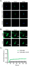
Figure 5.. PyS2–488 binds P. aeruginosa and achieves periplasmic localization.
PAO1 and PAO1 pretreated with 100 μM CCCP were stained with 1 μM PyS2–488 (see Methods) and examined with fluorescence microscopy techniques. (A) Super-resolution SIM confirmed periplasmic and outer membrane localization of PyS2–488 (Scale bar = 3 μm, Excitation: 488 nm, Emission: 528 nm). (B) Confocal fluorescence microscopy demonstrated localization of PyS2–488 to the outer membrane and periplasm (Scale bar = 3 μm, Excitation: 488 nm, Emission: 525/40 nm). In both microscopy experiments SYTO 40 Blue was used as a nonspecific bacterial stain to localize and focus on the bacteria (Excitation: 405 nm, Emission: 435 nm for SIM and 447/60 nm for confocal). (C) FRAP analysis illustrates PyS2–488 achieves periplasmic localization as a result of CCCP-dependent recovery.

References

    1. Kent SBH Novel Protein Science Enabled by Total Chemical Synthesis. Protein Sci. 2019, 28 (2), 313–328. - PMC - PubMed
    1. Rosano GL; Ceccarelli EA Recombinant Protein Expression in Escherichia Coli: Advances and Challenges. Front. Microbiol 2014, 5 (APR), 172. - PMC - PubMed
    1. Nilsson BL; Soellner MB; Raines RT Chemical Synthesis of Proteins. Annu. Rev. Biophys. Biomol. Struct 2005, 34 (1), 91–118. - PMC - PubMed
    1. Tan Y; Wu H; Wei T; Li X Chemical Protein Synthesis: Advances, Challenges, and Outlooks. J. Am. Chem. Soc 2020, 142 (48), 20288–20298. - PubMed
    1. Kulkarni SS; Sayers J; Premdjee B; Payne RJ Rapid and Efficient Protein Synthesis through Expansion of the Native Chemical Ligation Concept. Nat. Rev. Chem 2018, 2 (4), 0122.

Publication types