. 2023 Feb 1;4(2):150-161.
doi: 10.34067/KID.0002942022. Epub 2022 Oct 18.

Cardiac Manifestations in Patients with Autosomal Dominant Polycystic Kidney Disease (ADPKD): A Single-Center Study

Affiliations

Cardiac Manifestations in Patients with Autosomal Dominant Polycystic Kidney Disease (ADPKD): A Single-Center Study

Sita Arjune et al. Kidney360. .

Abstract

Key Points:

  1. Cardiovascular disease—a key driver of morbidity in CKD—is common in patients with autosomal dominant polycystic kidney disease (ADPKD).

  2. Pathologic echocardiography findings, including valvular defects, aortic root dilation, and hypertrophy, are found in most patients with ADPKD.

  3. These findings correlate with parameters indicating disease progression in ADPKD. Echocardiography should be offered to all patients with ADPKD.

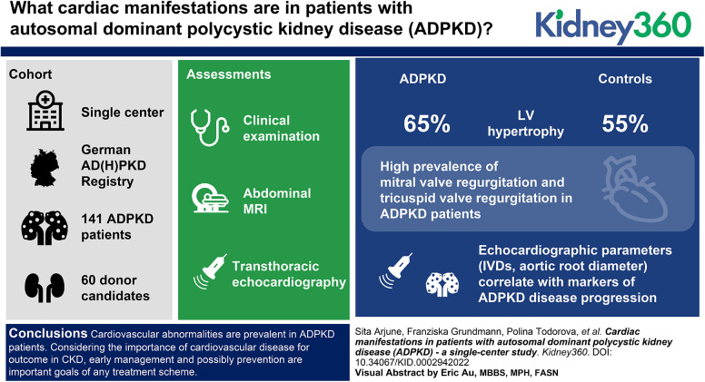
Background: ADPKD is the most common monogenetic kidney disease and results in kidney failure in >75% of affected individuals. As a systemic disorder, ADPKD is associated with a variety of extrarenal manifestations, including cardiac manifestations, that affect the majority of patients. We characterized the cardiac involvement in patients with ADPKD from the German AD(H)PKD registry and compared them with kidney donor candidates as controls.

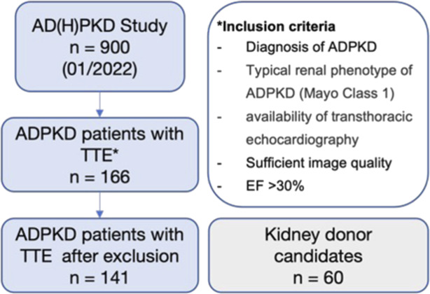
Methods: In this single-center cohort study, we evaluated 141 patients with ADPKD (44.17±11.23 years) from the German AD(H)PKD registry and 60 kidney donor candidates (55.08±10.21 years). All patients underwent clinical examination, abdominal MRI, and transthoracic echocardiography.

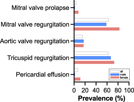
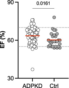
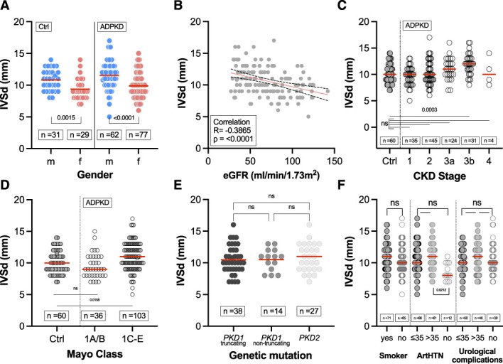
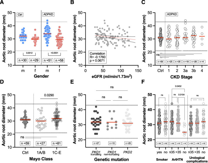
Results: Of the patients with ADPKD, 65% showed hypertrophy of the left ventricle (as defined by an end-diastolic interventricular septal wall thickness [IVSd] >10 mm) compared with 55% in control patients. Mitral regurgitation was the most common finding among 54% of patients with ADPKD who exhibited valvular dysfunction, albeit mild in most patients. Interestingly, left ventricular ejection fraction (LV-EF) differed significantly between both groups, with higher values in patients with ADPKD (64%±6% versus 60%±6%), whereas other parameters, including IVSd, left ventricular end-diastolic diameter (LVEDD), tricuspid annular plane systolic excursion (TAPSE), and pressure gradients across the aortic and tricuspid valve were similar between groups. Correlations of echocardiographic parameters with markers of disease progression revealed statistically significant associations for aortic root diameter (P=0.01), the pressure gradient across the aortic valve (AV dPmax; P=0.0003), and IVSd (P=0.0001), indicating rapid kidney disease progression may also be associated with cardiac findings.

Conclusion: Cardiovascular abnormalities are prevalent in patients with ADPKD. Considering the importance of cardiovascular disease for outcomes in CKD, early management and possibly prevention are important goals of any treatment scheme. Consequently, echocardiography should be offered to all patients with ADPKD in routine management.


Conflict of interest statement

R.-U. Müller reports having consultancy agreements with, and receiving honoraria from, Alnylam and Sanofi; having ownership interest in Bayer, ChemoCentryx, Novartis, Pfizer, Roche, and Santa Barbara Nutrients; serving as chair of the working group "Genes and Kidney" of the European Renal Association (ERA) and member of the working group on AD dysplasias of the European Rare Kidney Disease Reference Network (ERKNet), on the editorial board of Kidney and Dialysis, and on the scientific advisory board of Santa Barbara Nutrients; and receiving research funding from Otsuka Pharmaceuticals and Thermo Fisher Scientific (all research funding was paid to the employer, Department II of Internal Medicine). R. Pfister reports receiving honoraria from Abbot Vascular, AstraZeneca, Bayer, Boehringer Ingelheim, Edwards Lifesciences, Novartis, Occlutech, and Pfizer; having consultancy agreements with Edwards Lifesciences and Occlutech; serving in an advisory or leadership role for Occlutech; and receiving research funding from Pfizer. H. ten Freyhaus reports receiving honoraria from Actelion, Alnylam, Bristol Myers Squibb, CTI, Diaplan, FomF, GlaxoSmithKline, Guerbet, KelCon, Lilly, Medtronic, medupdate, Novartis, Pfizer, Sanofi-Aventis, Servier, TomTec, and Volcano; having consultancy agreements with Alnylam, AstraZeneca, Bayer, GE, and Janssen-Cilag; and receiving research funding from Alnylam and Ionis. P. Todorova reports receiving travel and accommodation costs from Alexion, Astellas, Bristol Myers Squibb, Chemocentryx, Chiesi, Novartis, Omeros, and Otsuka. All remaining authors have nothing to disclose.

Figures

None
Graphical abstract
Figure 1
Figure 1
Study flowchart illustrating patient flow and inclusion criteria. ADPKD, autosomal dominant polycystic kidney disease; EF, ejection fraction; TTE, transthoracic echocardiography.
Figure 2.
Figure 2.
Prevalence of cardiac valve defects and pericardial effusion in patients with ADPKD.
Figure 3.
Figure 3.
Comparison of left ventricular EF of patients with ADPKD versus controls. Mean values for patients with ADPKD (64%) and controls (Ctrl; 60%) are indicated by red lines.
Figure 4.
Figure 4.
Correlation of htTKV and echocardiographic parameters. Pearson correlation was conducted after the D’Agostino and Pearson test revealed parametric data distribution. The red line indicates simple linear regressions; black dotted lines indicate the 95% CI. AV, aortic valve; dPmax, pressure gradient; IVSd, end-diastolic interventricular septal wall thickness; LVEDD, left ventricular end-diastolic diameter; htTKV, height-adjusted TKV; TAPSE, tricuspid annular plane systolic excursion; TV, tricuspid valve.
Figure 5.
Figure 5.
Association of end-diastolic interventricular septal wall thickness (IVSd) with clinical characteristics. (A) Sex distribution of IVSd in patients with ADPKD (mean±SD of 11.55±2.18 mm for men and 9.88±1.97 mm for women) and controls (mean±SD of 10.82±1.65 mm for men and 9.38±1.70 mm for women). (B) Pearson correlation of IVSd and eGFR in patients with ADPKD. (C) IVSd according to CKD stages 1–4 in patients with ADPKD (mean±SD of 9.66±1.78 mm for CKD1, 10.27±2.30 mm for CKD2, 11.04±2.12 mm for CKD3a, 11.93±1.98 mm for CKD3b, 10.50±2.65 mm for CKD4) versus controls (mean±SD of 10.13±1.81 mm). (D) IVSd according to Mayo classification (1A/B [mean±SD of 9.53±1.34 mm] versus 1C/D/E [mean±SD of 11.01±2.19 mm]) in patients with ADPKD versus controls (mean±SD of 10.13±1.81 mm). (E) IVSd in patients with ADPKD among respective gene products (PKD1 and PKD2). Truncating mutations included nonsense, frameshift, splicing mutations, and large rearrangements, whereas nontruncating mutations included missense mutations and in-frame short deletions and insertions. (F) IVSd for smokers and relevant comorbidities (for ArtHTN: mean±SD of 10.63±2.28 mm for ≤35 years, 11.02±2.86 mm for >35, 8.58±1.38 mm for no ArtHTN). Horizontal red lines indicate mean values. ArtHTN, arterial hypertension.
Figure 6.
Figure 6.
Correlation of IVSd with age in (A) patients with ADPKD and (B) controls. Pearson correlation was conducted after D’Agostino and Pearson test revealed parametric data distribution. The red line indicates simple linear regressions; black dotted lines indicate the 95% CI.
Figure 7
Figure 7
Association of aortic root diameter with clinical characteristics. (A) Sex distribution of aortic root diameter in patients with ADPKD (mean±SD of 33.96±4.06 mm for men and 29.45±3.52 mm for women) and controls (mean±SD of 32.13±2.75 mm for men and 29.90±2.42 mm for women) by sex. (B) Pearson correlation of aortic root diameter and eGFR in patients with ADPKD. (C) Aortic root diameter according to CKD stages 1–4 in patients with ADPKD (mean±SD of 31.40±4.92 mm for CKD1, 30.49±4.17 mm for CKD2, 32.79±4.59 mm for CKD3a, 32.15±3.79 mm for CKD3b, 32.25±5.56 mm for CKD4) versus controls (mean±SD of 31.03±2.74 mm). (D) Aortic root diameter according to Mayo classification (1A/B [mean±SD of 30.04±4.78mm] versus 1C/D/E [mean±SD of 32.15±4.12 mm]) in patients with ADPKD versus controls (mean±SD of 31.03±2.74 mm). (E) Aortic root diameter in patients with ADPKD among respective gene products (PKD1 and PKD2). Truncating mutations included nonsense, frameshift, splicing mutations, and large rearrangements, whereas nontruncating mutations included missense mutations and in-frame short deletions and insertions. (F) Aortic root diameter according to (ever) smokers and relevant comorbidities (for ArtHTN, mean±SD of 31.58±4.34 mm for ≤35 years, 32.33±4.16 mm for >35, 26.50±3.024 mm for no ArtHTN). Horizontal red lines indicate mean values. ArtHTN, arterial hypertension.

Comment in

  • Defining Cardiac Dysfunction in ADPKD.
    Kuo IY. Kuo IY. Kidney360. 2023 Feb 1;4(2):126-127. doi: 10.34067/KID.0000000000000066. Kidney360. 2023. PMID: 36821601 Free PMC article. No abstract available.

References

    1. Chapman AB Devuyst O Eckardt KU, et al. Conference Participants . Autosomal-dominant polycystic kidney disease (ADPKD): Executive summary from a Kidney Disease: Improving Global Outcomes (KDIGO) Controversies Conference. Kidney Int. 2015;88:17-27. 10.1038/ki.2015.59 - DOI - PMC - PubMed
    1. Müller RU, Benzing T. Cystic kidney diseases from the adult nephrologist’s point of view. Front Pediatr. 2018;6:65. doi:10.3389/fped.2018.00065 - DOI - PMC - PubMed
    1. Torres VE, Harris PC, Pirson Y. Autosomal dominant polycystic kidney disease. Lancet. 2007;369:1287-1301. doi:10.1016/S0140-6736(07)60601-1 - DOI - PubMed
    1. Neumann HP Jilg C Bacher J, et al. Else-Kroener-Fresenius-ADPKD-Registry . Epidemiology of autosomal-dominant polycystic kidney disease: An in-depth clinical study for south-western Germany. Nephrol Dial Transplant. 2013;28:1472-1487. doi:10.1093/ndt/gfs551 - DOI - PubMed
    1. Kataoka H Fukuoka H Makabe S, et al. . Prediction of renal prognosis in patients with autosomal dominant polycystic kidney disease using PKD1/PKD2 mutations. J Clin Med. 2020;9:146. doi:10.3390/jcm9010146 - DOI - PMC - PubMed

Publication types