

Published Erratum
doi: 10.1371/journal.pbio.3002027.
eCollection 2023 Feb.
Correction: The Naturally Processed CD95L Elicits a c-Yes/Calcium/PI3K-Driven Cell Migration Pathway
- PMID: 36821824
- PMCID: PMC9949890
- DOI: 10.1371/journal.pbio.3002027
Item in Clipboard
Published Erratum
Correction: The Naturally Processed CD95L Elicits a c-Yes/Calcium/PI3K-Driven Cell Migration Pathway
PLoS Biol.
.
Abstract
[This corrects the article DOI: 10.1371/journal.pbio.1001090.].
Copyright: © 2023 Tauzin et al. This is an open access article distributed under the terms of the Creative Commons Attribution License, which permits unrestricted use, distribution, and reproduction in any medium, provided the original author and source are credited.
Figures
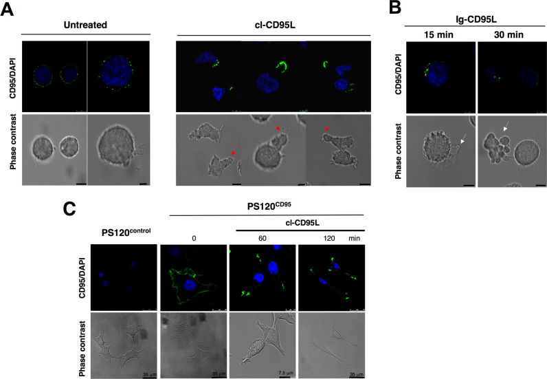
(A) Activated (PHA/IL2)-peripheral blood T-lymphocytes (PBLs) from healthy individuals were incubated with control medium (untreated condition corresponding to the incubation with supernatant of pcDNA3.1(+)-transfected HEK cells) or 100 ng/ml of cl-CD95L for 30 min. Cell morphology was followed using phase contrast microscopy and CD95 was stained using a mouse anti-CD95 mAb (APO1-3) followed by a goat Alexa488-coupled anti-mouse IgG mAb. Red arrows depict emitted pseudopods (Bars = 5 mm). (B) PHA/IL2-activated PBLs were incubated with the cytotoxic Ig-CD95L for indicated times, and cells were analyzed as described in (A). White arrows depict blebs emitted by apoptotic cells (bars = 5 mm). (C) The fibroblastic cell line PS120 devoid of endogenous CD95 (wild type NHE1-reconstituted PS120control) or reconstituted with human wild type CD95 (PS120CD95) were untreated or treated with 100 ng/ml of cleaved CD95L for indicated times, and cell shape and CD95 distribution were analyzed. The “0” condition indicates cells analyzed before the addition of 100 ng/mL of cl-CD95L.
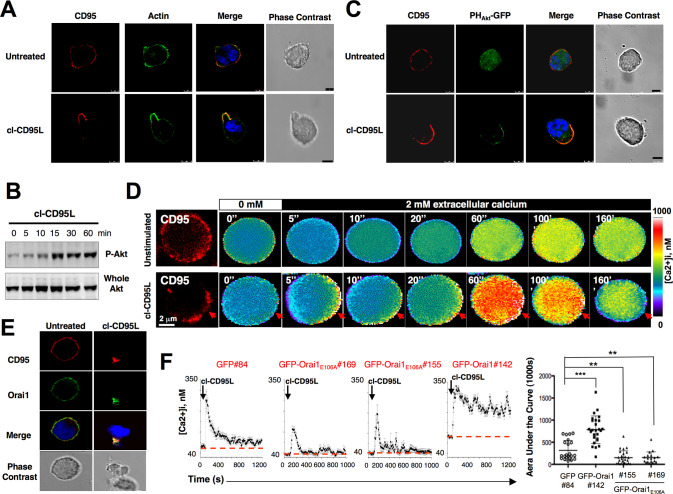
(A) H9 T-cells were transiently transfected with the actin marker Lifeact-GFP. Living cells were harvested (Ficoll) and treated for 30 min with 100 ng/ml of cleaved CD95L or with control medium (supernatant of pcDNA3.1(+)-transfected HEK cells—untreated). Cells were fixed, and CD95 was stained using anti-CD95 mAb (APO1-3) and revealed with the secondary Alexa555-coupled Goat anti-mouse antibody (red). Nuclei were stained using DAPI (blue). Bars = 5 mm. Images were acquired with an ApoPLAN 63× objective. (B) The leukemic T-cell line H9 was incubated with 100 ng/ml of the naturally processed CD95L for indicated times. Cells were lysed, and for each lane, 100 mg of protein were loaded. Proteins were resolved by SDS-PAGE and anti-whole Akt and anti-Akt phosphorylation (serine473) immunoblots were performed. Akt phosphorylation stands for its activation. (C) The pleckstrin homology (PH) domain of Akt binds PIP3 produced by PI3K activation. As a consequence, the probe PHAkt-GFP stains PIP3. H9 T-cells were transiently transfected with the PHAkt-GFP construct (green). Living cells were isolated (Ficoll) and incubated with 100 ng/ml of the naturally processed CD95L (cl-CD95L) or with control medium (supernatant from pcDNA3.1(+)-transfected HEK cells—untreated) for 30 min. Cells were fixed, and CD95 was stained as previously mentioned (red). Pictures were taken by confocal microscopy. Cell morphology was followed using differential interference contrast microscopy. Nuclei were stained with DAPI (blue). Bars = 5 mm. (D) Calcium measurement in single cell. Jurkat T-cells were loaded with the permeant calcium probe Fura-2AM, and in parallel, CD95 was followed using an anti-CD95 mAb and an Alexa555-coupled goat anti-mouse mAb as described in Materials and Methods. Cells were bathed in a Ca2+-free medium and pre-incubated (lower panel; cl-CD95L) or not (upper panel; supernatant from pcDNA3.1(+)-transfected HEK cells—untreated)) with cl-CD95L (100 ng/ml). 2 mM Ca2+ was added when indicated by the black-filled rectangle. Ratio images (F340 nm/F380 nm) were taken every 5 s, and the images shown correspond to the indicated period of time. Grey level intensities were translated to false colors according to the colors scale shown at the right, and [Ca2+]i was estimated from the ratio values and calibration experiments as described in Materials and Methods. Red arrows indicate the CD95-CAP. (E) Jurkat T-cells were treated with 100 ng/ml of cl-CD95L or with control medium (supernatant from pcDNA3.1(+)-transfected HEK cells—untreated) for 60 min. Cells were fixed and Orai1 and CD95 were stained as described in Materials and Methods. Cell morphology was analyzed using phase contrast microscopy. (F) Left panels: GFP-, GFP-Orai1, or GFP-Orai1E106A-expressing Jurkat T-cells were loaded with 1 mM of the calcium probe Fura-2AM for 30 min at RT. Cells were bathed at 37°C in a medium containing 2 mM [Ca2+]e and then treated with 100 ng/ml of cl-CD95L (black arrow). GFP#84 corresponded to control cells expressing GFP. GFP-Orai1E106A#155 and #169 corresponded to two independently isolated clones expressing the non-functional CRAC channel. GFP-Orai1#142 corresponded to Jurkat cells overexpressing human Orai1. Values were recorded every 10 s. Right panel: For each experiment, the area under the curve (AUC) was measured for 1,000 s, and the statistical analyses of the AUC values were performed for indicated cells using non-parametric two-tailed Mann-Whitney tests. ** and *** indicate p values≤0.01 and 0.001, respectively.
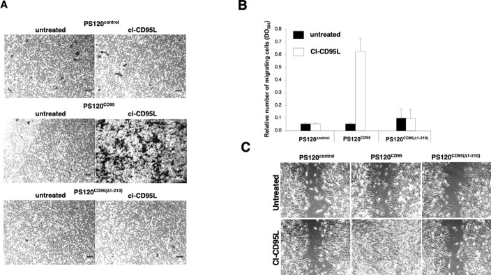
(A) The CD95-deficient PS120 (wild type NHE1-reconstituted PS120control) and its counterparts expressing human wild type CD95 or DD-truncated CD95 were seeded in a Boyden chamber in the presence of cl-CD95L (100 ng/ml) or with control medium (supernatant from pcDNA3.1(+)-transfected HEK cells—Untreated) and incubated for 24 h. The filter was removed, the upper side containing the non-migrating cells was wiped out with cotton-tipped swabs and migrating cells in the opposite side of the filter were fixed with methanol and stained (Giemsa). For each experiment, five pictures of random fields were taken, and a representative picture was depicted (Bars = 70 μm). (B) Cells were treated as described in (A). To quantify cell motility, Giemsa-stained migrating cells from the lower side of the membrane were lyzed and absorbance was measured at 560 nm. (C) Wound healing assay, a confluent monolayer of the indicated cells was “wounded” with a tip and then cells were incubated for 24 h in the presence of 100 ng/ml of cl-CD95L or a control medium (supernatant of pcDNA3.1(+)-transfected HEK cell) (untreated) and pictures were acquired (Bars = 50 μm). Pictures are representative of 5 independently performed experiments.
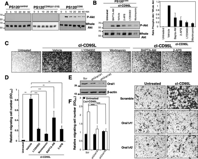
(A) The CD95-deficient fibroblastic cell line PS120 transfected with empty vector (wild type NHE1-reconstituted PS120control) or reconstituted with either human wild type CD95 (PS120CD95) or a death domain-truncated CD95 (PS120CD95(Δ1–210)) was incubated with 100 ng/ml of cleaved CD95L for indicated times. Cells were harvested, lyzed, and 100 μg of protein was loaded per lane. Proteins were resolved in a SDS-PAGE and immunoblots were performed. Phosphorylation of the serine473 on Akt indicates activation of the kinase. Whole Akt is added as loading control. The PS120 immunoblots shown in (A) have been partly reused in [3]. (B) Left panels: PS120CD95 cells were pre-incubated with non-cytotoxic and non-cytostatic concentration of PI3K inhibitors (2.5 μM of wortmannin and 5 μM of LY294002), the cell permeant chelator of calcium BAPTA-AM (5 μM), the inhibitor of IP3-R and SOC channels 2-APB (20 μM) or DMSO (vehicle). Cells were then untreated (control medium from pcDNA3.1(+)-transfected HEK cells) or treated for 5 min with 100 ng/ml of cl-CD95L and lysed, 50 μg/ml of protein was loaded per lane and indicated immunoblots were performed. Right panel: the densitometry analyses of the immunoblot bands were performed using ImageJ software and the histograms depict Phospho-Akt/whole Akt ratios. (C) Cell migration of PS120CD95 was assessed using the Boyden chamber assay. Cells were pre-incubated with non-cytotoxic and non-cytostatic concentrations of PI3K inhibitors (2.5 μM of Wortmannin and 5 μM of LY294002), the cell permeant chelator of calcium BAPTA-AM (5 μM), the inhibitor of SOC channels 2-APB (20 μM) or DMSO (vehicle) for 30 min and then stimulated with 100 ng/ml of cl-CD95L or a control medium (supernatant from pcDNA3.1(+)-transfected HEK cell—untreated) for 24 h. For each experiment, 10 pictures of the migrating cells were taken, and a representative picture was depicted (Bars = 50 μm). (D) Cells were treated as described in (C). To quantitatively measure cell motility, Giemsa-stained migrating cells from the lower side of the membrane were lyzed, and absorbance was measured at a wavelength of 560 nm. Values represent means and SD of three independently performed experiments. **p≤0.01 and ***p≤0.001 as calculated using two-tailed non-parametric Mann-Whitney test. (E) The silencing effect of the Orai1-targeting shRNAmir-pGIPZ vectors was analyzed by immunoblot in lentiviral-transduced HEK cells. 48 h after transduction, cells were lysed and 100 mg of lysates were loaded per line. β-actin serves as a loading control. (F) Cell migration of indicated shRNAmir-transduced HEK cells was assessed using Boyden chamber assay. Right panels: HEK cells were transduced with scrambled or Orai1-targeting ShRNA, and 48 h later, cells were incubated for 24 h in the presence of 100 ng/ml of cl-CD95L or untreated (supernatant of pcDNA3.1(+)-transfected HEK cells). Migrating cells were fixed with methanol and stained by Giemsa. For each experiment, five pictures of random fields were taken, and a representative picture was depicted (Bars = 70 mm). Left panel: To quantitatively measure cell motility, Giemsa-stained migrating cells from the lower side of the membrane were lyzed, and absorbance was measured at a wavelength of 560 nm. Values represent means and SD of three independently performed experiments. *** p≤0.001 as calculated using two-tailed non-parametric Mann-Whitney test.
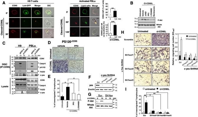
(A) Left panels: The leukemic cell line H9 was transiently transfected with Lck-GFP. 24 h after transfection, living cells were treated or untreated for the indicated times in the presence of cl-CD95L (100 ng/ml). 0 min (0’) condition corresponds to cells analyzed prior to the addition of 100 ng/mL of cl-CD95L. CD95 was stained using a mouse anti-CD95 mAb and a goat Alexa555-coupled anti-mouse IgG mAb. Right panels: Activated PBLs were incubated for the indicated times with 100 ng/ml of cl-CD95L, and then cells were fixed. Lipid rafts were stained using Alexa488-cholera toxin B subunit and CD95 was followed as previously mentioned. Images were acquired with a confocal microscope with a 63× objective. 0 min (0’) condition corresponds to cells analyzed prior to the addition of 100 ng/mL of cl-CD95L. Cell morphology was followed using phase contrast microscopy (Bars = 7.5 mm). The percentage of T-cells displaying a CD95 cluster was assessed (300 cells counted for each condition). Among the activated T-lymphocytes showing CD95 cluster, the amount of CD95-CAP co-localized with lipid rafts was assessed. (B) H9 T-cells were pre-incubated for 30 min with DMSO (vehicle), 10 mM of PP2, 5 mM of LY294002 (LY), 2.5 mM of wortmannin (Wort), or 40 mM of zVAD-fmk (zV) and then treated for 15 min with 100 ng/ml of cl-CD95L. Untreated condition corresponds to cells incubated with supernatant of pcDNA3.1(+)-transfected HEK cells. Cells were lyzed and Akt phosphorylation (S473) and whole Akt were assessed by immunoblots. (C) The H9 T-cell line (left panels) and activated PBLs (right panels) were incubated for indicated times with 100 ng/ml of cl-CD95L or APO1-3. APO1-3 is an agonistic anti-CD95 mAb incubated for 15 min with indicated cells to activate CD95 and trigger DISC formation. Then, cells were lyzed and CD95 was immunoprecipitated. The 0 min condition corresponds to unstimulated cells directly lyzed to next immunoprecipitate CD95. APO1-3 also serves for the immunoprecipitation step of CD95 (see Materials and Methods). The immune complex was resolved in a 10% SDS-PAGE and indicated immunoblots were performed. Total lysates were depicted to confirm that the same amount of protein was present for each immunoprecipitation. The black stars represent heavy chains of Ig. (D) Boyden chamber assays were performed as described in Materials and Methods. PS120CD95 (All PS120 cells have been reconstituted with wild type NHE1 (see Materials and Methods) were pre-incubated with or without a non-cytotoxic and non-cytostatic concentration of PP2 (10 mM) or DMSO (vehicle) for 30 min and then treated (cl-CD95L) or untreated (Untreated condition corresponds to cells incubated with supernatant of pcDNA3.1(+)-transfected HEK cells) for 24 h with 100 ng/ml of cl-CD95L. Data are representative of three independently performed experiments. The vehicle conditions in the presence or absence of cl-CD95L also serve in S9B Fig to decipher the effect of zVAD-fmk. (E) Activated PBLs were pre-incubated with 10 mM of the src inhibitor PP2, 5 mM of the PI3K inhibitor LY294002 (LY) or DMSO (vehicle) and then incubated in the presence of cl-CD95L (100 ng/ml) for 24 h. Untreated cells correspond to activated PBLs incubated for 24h with supernatant of pcDNA3.1(+)-transfected HEK cells. Then, endothelial transmigration of PBLs was assessed as described in Materials and Methods. (F) The silencing effect of the c-yes-targeting shRNAmir-pGIPZ vectors was analyzed by immunoblot in lentiviral-transduced PS120CD95 cells. 72 h after transduction, cells were lysed and 100 mg of lysate was loaded per line. β-actin serves as loading control. (G) The T-cell line H9 was transduced with scramble (scr) or c-yes (shYes#3)-targeting shRNAmir-containing lentivirus. 72 h after transduction, living cells were harvested and green cells were sorted by flow cytometry using FACSAria. Cells were immediately stimulated with cl-CD95L (100 ng/ml) (15 min) or left untreated (0 min), and the amounts of Akt phosphorylation (S473) were assessed by immunoblot. (H) Cell migration of indicated shRNAmir-transduced PS120CD95 was assessed using the Boyden chamber assay. Left panels: 72 h after transduction, cells were treated (cl-CD95L) or untreated (supernatant of pcDNA3.1(+)-transfected HEK cells) with 100 ng/ml of cl-CD95L for 24 h. For each experiment, five pictures of the migrating cells were taken, and a representative picture was depicted (Bars = 50 mm). Right panel: To quantitatively measure cell motility, Giemsa-stained migrating cells from the lower side of the membrane were lyzed and absorbance was measured at a wavelength of 560 nm. Values represent means and SD of three independently performed experiments. (I) H9 cells were transduced as mentioned in (G), and the green cells were incubated in the presence of cl-CD95L (100 ng/ml) or supernatant of pcDNA3.1(+)-transfected HEK cells (untreated) for 24 h. Then, the endothelial transmigration of the sh-RNA-expressing T-cells was assessed as described in Materials and Methods.
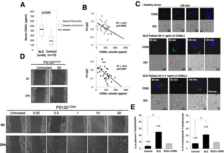
(A) By ELISA, the amounts of soluble CD95L in sera of healthy donors (control) and SLE patients were quantified. n stands for the number of SLE patients and healthy subjects. (B) Correlation between aggravation markers of the disease and the amount of soluble CD95L. (C) Activated PBLs were incubated for the indicated times with sera of healthy donors or SLE patients and the distribution of CD95 was analyzed by confocal microscopy. (D) All PS120 cells have been reconstituted with wild type NHE1 (see Materials and Methods). PS120control cells do not express CD95, while its counterpart PS120CD95 expresses a full length human CD95 (see Materials and Methods). A 24 h wound healing assay was performed. The fibroblastic cell line PS120control or its CD95-expressing counterpart PS120CD95 was grown to confluence. then the monolayer was “wounded” by scratching cells using a pipette tip. Using microscopy, the migration was monitored for 24 h with the indicated concentrations of cl-CD95L (ng/mL) as indicated in Materials and Methods. The untreated condition corresponds to cells incubated with a control medium (supernatant of pcDNA3.1(+)-transfected HEK cells). Pictures are representative of 3 independently performed experiments. (E) Activated PBLs were incubated in the presence of sera from SLE patients (n = 3) or healthy donors (n = 6), and then, adhesion to endothelial cells (left panel) and endothelial transmigration (right panel) of PBLs were assessed as described in Materials and Methods. Where indicated, activated PBLs were preincubated 30 min with the antagonist anti-CD95 mAb ZB4 (α-CD95). ZB4 was maintained in the culture at 10 μg/ml.
Erratum for
-
The naturally processed CD95L elicits a c-yes/calcium/PI3K-driven cell migration pathway.PLoS Biol. 2011 Jun;9(6):e1001090. doi: 10.1371/journal.pbio.1001090. Epub 2011 Jun 21. PLoS Biol. 2011. PMID: 21713032 Free PMC article.
-
Correction: The Naturally Processed CD95L Elicits a c-Yes/Calcium/PI3K-Driven Cell Migration Pathway.PLoS Biol. 2019 Oct 8;17(10):e3000521. doi: 10.1371/journal.pbio.3000521. eCollection 2019 Oct. PLoS Biol. 2019. PMID: 31593568 Free PMC article.
References
-
- Billottet C, Banerjee L, Vanhaesebroeck B, Khwaja A. Inhibition of class I phosphoinositide 3-kinase activity impairs proliferation and triggers apoptosis in acute promyelocytic leukemia without affecting atra-induced differentiation. Cancer Res. 2009;69(3):1027–36. Epub 2009/01/30. doi: 10.1158/0008-5472.CAN-08-2608 . - DOI - PubMed
-
- Hayakawa M, Kaizawa H, Moritomo H, Koizumi T, Ohishi T, Okada M, et al.. Synthesis and biological evaluation of 4-morpholino-2-phenylquinazolines and related derivatives as novel PI3 kinase p110alpha inhibitors. Bioorg Med Chem. 2006;14(20):6847–58. Epub 2006/07/14. doi: 10.1016/j.bmc.2006.06.046 . - DOI - PubMed
Publication types
LinkOut - more resources
Full Text Sources
