Review
. 2023 Apr:79:102549.
doi: 10.1016/j.sbi.2023.102549. Epub 2023 Feb 21.

MicroED in drug discovery

Affiliations
Review

MicroED in drug discovery

Emma Danelius et al. Curr Opin Struct Biol. 2023 Apr.

Abstract

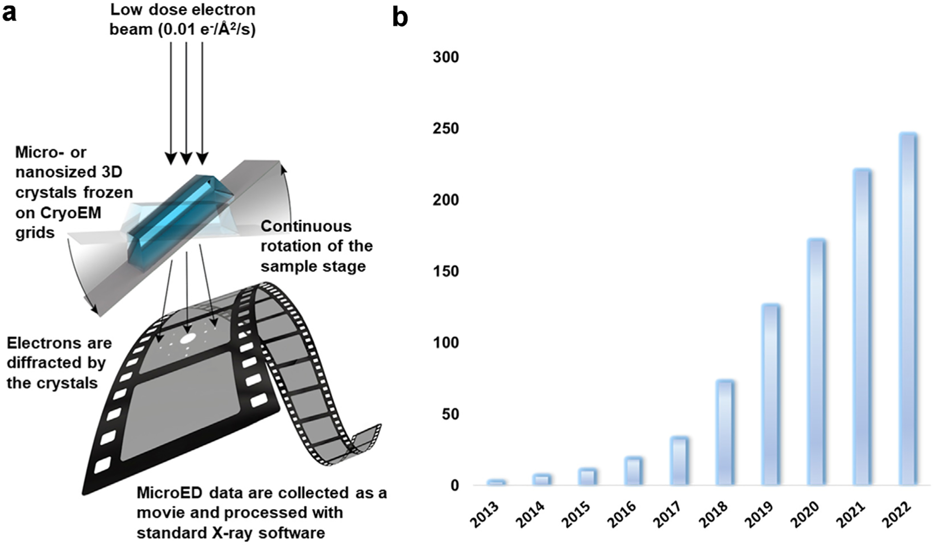
The cryo-electron microscopy (cryo-EM) method microcrystal electron diffraction (MicroED) was initially described in 2013 and has recently gained attention as an emerging technique for research in drug discovery. As compared to other methods in structural biology, MicroED provides many advantages deriving from the use of nanocrystalline material for the investigations. Here, we review the recent advancements in the field of MicroED and show important examples of small molecule, peptide and protein structures that has contributed to the current development of this method as an important tool for drug discovery.

Keywords: Cryo-EM; MicroED; Nanocrystals; Protein-ligand structures; Small molecule structures.


Conflict of interest statement

Declaration of competing interest The authors declare that they have no known competing financial interests or personal relationships that could have appeared to influence the work reported in this paper.

Figures

Figure 1
Figure 1
In MicroED, three-dimensional micro or nanosized crystals are continuously rotated in the electron beam in the TEM, and the data are collected on a high-speed camera as a movie. The data can be processed using the well-established X-ray diffraction programs. (B) Since its initial demonstration in 2013, the number of published MicroED structures have increased exponentially. The graph shows the total accumulated number of MicroED structures deposited in the pdb and CCDC databases as of the preparation of this figure.
Figure 2
Figure 2
MicroED of small molecules: (a) MicroED workflow of small molecule structure determination directly from powder, without any prior crystallization step.(b) Recent pharmaceuticals and natural product structures determined by MicroED. (c) MicroED structure of 24DHPA with density map contoured at 1σ.(d) Structures of the original and revised assignment of the lomaiviticins core.
Figure 3
Figure 3
Examples of ligand-bound protein structures solved by MicroED: a. The HCA II–AZM binding site from a 2.5 Å resolution MicroED structure [29]. b. The3.2 Å structure of catalase solved by MicroED, showing the heme binding site [37]. c. The MicroED structure of the CTD-SP1 fragment of HIV-1 Gag with bound maturation inhibitor bevirimat [38]. d. The 2.5 Å MicroED structure of a protoglobin reactive carbene intermediate at the heme binding site [39]. e. MicroED structure of the human adenosine receptor at 2.8 Å, showing the binding site of the antagonist ZMA (grey) as well as several cholesterols binding to the membrane helices (pink) [35].
Figure 4
Figure 4
Phasing in MicroED: a. The 0.87 Å resolution structure of lysozyme was determined with ab initio phasing. This sub-Ångstrom structure showed density for hydrogens (displayed in green) as well revealed the histidine 15 to be charged, as shown here in the close-up of this residue with the corresponding hydrogen bonds to alanine 11 and threonine 89 [41]. b. The novel structure of a protoglobin Aeropyrum pernix protoglobin engineered for carbene transfer reactions in asymmetric synthesis. No wild-type homologue was available and the structure was determined using an AlphaFold2-predicted structure for phasing of the MicroED data [42].

References

    1. Nannenga BL: MicroED methodology and development. Structural Dynamics 2020, 7, 014304. - PMC - PubMed
    1. Christensen J, Horton PN, Bury CS, Dickerson JL, Taberman H, Garman EF, Coles S: Radiation damage in small-molecule crystallography: fact not fiction. IUCrJ 2019, 6:703–713. - PMC - PubMed
    1. Shi D, Nannenga BL, Iadanza MG, Gonen T: Three-dimensional electron crystallography of protein microcrystals. Elife 2013, 2, e01345. - PMC - PubMed
    1. Nannenga BL, Shi D, Leslie AGW, Gonen T: High-resolution structure determination by continuous-rotation data collection in MicroED. Nat Methods 2014, 11:927–930. - PMC - PubMed
    1. van Genderen E, Clabbers MT, Das PP, Stewart A, Nederlof I, Barentsen KC, Portillo Q, Pannu NS, Nicolopoulos S, Gruene T, Abrahams JP: Ab initio structure determination of nanocrystals of organic pharmaceutical compounds by electron diffraction at room temperature using a Timepix quantum area direct electron detector. Acta Crystallogr A 2016, 72:236–242. - PMC - PubMed

Publication types