. 2023 Apr 19;111(8):1205-1221.e9.
doi: 10.1016/j.neuron.2023.01.022. Epub 2023 Feb 22.

DNA-initiated epigenetic cascades driven by C9orf72 hexanucleotide repeat

Affiliations

DNA-initiated epigenetic cascades driven by C9orf72 hexanucleotide repeat

Yang Liu et al. Neuron. .

Erratum in

Abstract

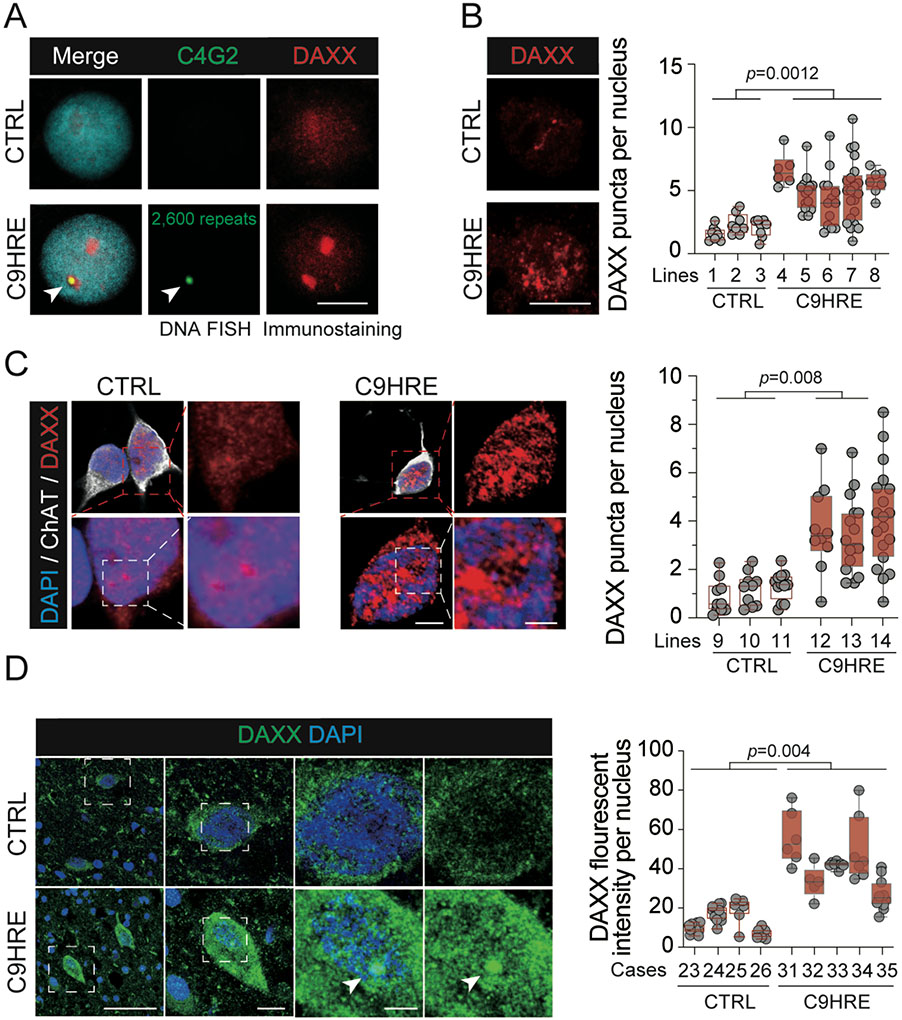
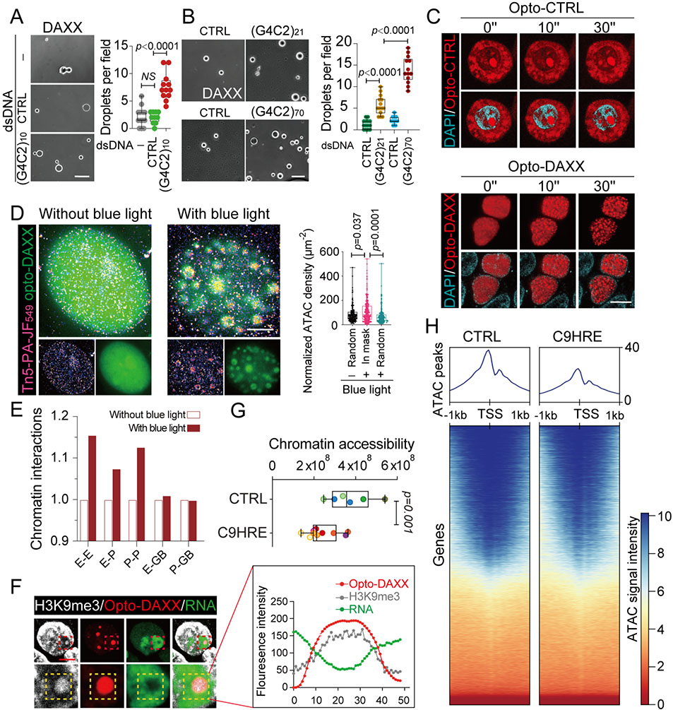
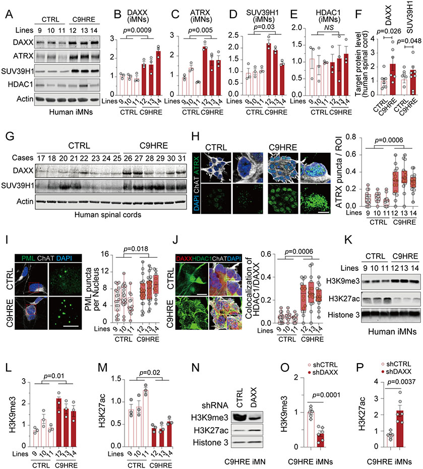
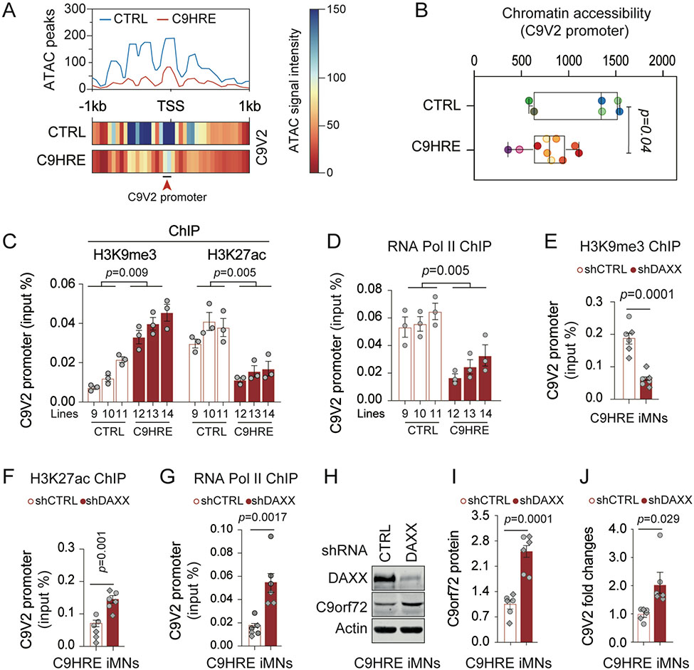
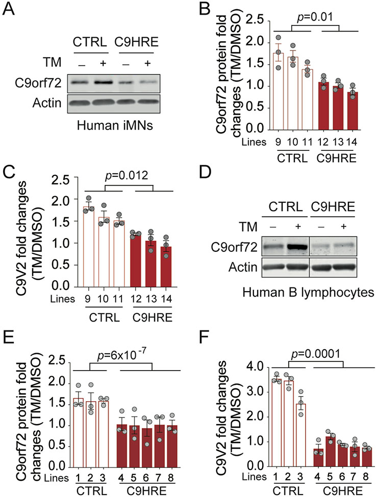
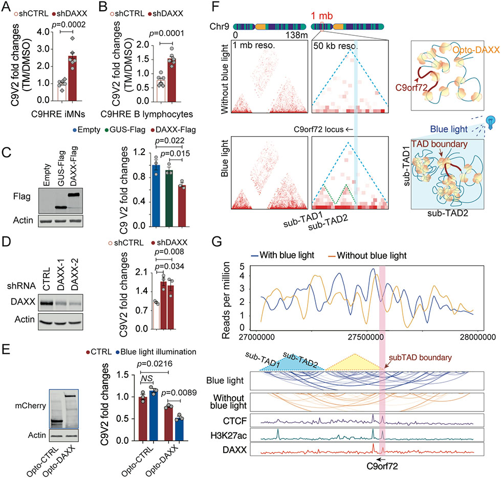
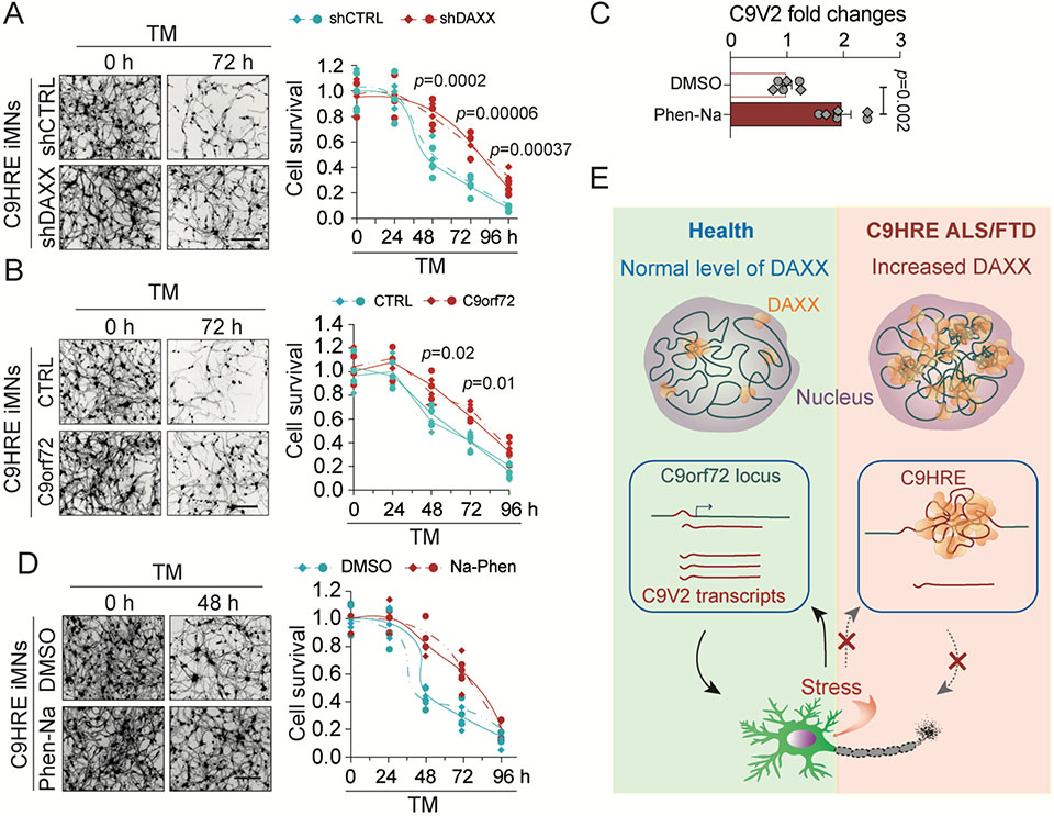
The C9orf72 hexanucleotide repeat expansion (HRE) is the most frequent genetic cause of the neurodegenerative diseases amyotrophic lateral sclerosis (ALS) and frontotemporal dementia (FTD). Here, we describe the pathogenic cascades that are initiated by the C9orf72 HRE DNA. The HRE DNA binds to its protein partner DAXX and promotes its liquid-liquid phase separation, which is capable of reorganizing genomic structures. An HRE-dependent nuclear accumulation of DAXX drives chromatin remodeling and epigenetic changes such as histone hypermethylation and hypoacetylation in patient cells. While regulating global gene expression, DAXX plays a key role in the suppression of basal and stress-inducible expression of C9orf72 via chromatin remodeling and epigenetic modifications of the promoter of the major C9orf72 transcript. Downregulation of DAXX or rebalancing the epigenetic modifications mitigates the stress-induced sensitivity of C9orf72-patient-derived motor neurons. These studies reveal a C9orf72 HRE DNA-dependent regulatory mechanism for both local and genomic architectural changes in the relevant diseases.

Keywords: ALS; C9orf72; DAXX; DNA; FTD; chromatin; epigenetic; neurodegeneration; phase separation; transcription.


Conflict of interest statement

Declaration of interests The authors declare no competing interests.

Figures

Figure 1.
Figure 1.. C9orf72 HRE-associated DAXX condensates in ALS patient cells.
(A) Immunoblotting of DAXX in the precipitates pulled down by a random or (G4C2)6 dsDNA biotin-labeled probe from nuclear fractions (n = 3 biological replicates). (B) Co-localization of the C9HRE DNA locus with one of the DAXX condensates. Representative images show DNA FISH analysis of the HRE locus using an Alexa Fluor 488-labeled (C4G2)4 ssDNA probe, with co-immunostaining for DAXX, for C9-ALS patient B lymphocytes harboring a $2,600 G4C2 repeat and control cells without the expanded repeat. A focal plane in which the repeat locus and one of the DAXX puncta co-localize (arrowhead) is shown. Scale bar, 10 mm. (C) Increased DAXX condensation in C9HRE ALS patient-derived motor neurons. Representative immunostaining images of DAXX in control and C9HRE motor neurons are shown. Ten fields containing 72–117 neurons from two independent slides for each cell line were examined (n = 3 independent pairs of iMN lines). Scale bars, 3 (left) and 1 (right) mm. (D) Increased immunostaining of DAXX in C9HRE patient spinal cord neurons. Representative DAXX immunostaining images are shown, and neurons are identified through their characteristic shapes. Each dot represents the average intensity of DAXX immunofluorescence per nucleus in a field of view (n = 5 C9HRE cases and 4 control cases). Scale bars, 100, 10, and 5 mm (from left to right). See also Figure S1.
Figure 2.
Figure 2.. Liquid-liquid phase separation of DAXX reorganizes chromatin topology and spatial transcription.
(A) The HRE dsDNA promotes the liquid-liquid phase separation of DAXX. Purified DAXX was incubated in LLPS buffer (1 M NaCl) without dsDNA or with (G4C2)10 dsDNA or a size-matched control dsDNA at room temperature for 0.5 h. 10–12 fields of view in each group were quantified and statistically analyzed. Scale bars, 10 mm. (B) The HRE repeat-length-dependent effects on the liquid-liquid phase separation of DAXX. Purified DAXX was incubated in LLPS buffer (0.5 M NaCl) with G4C2 dsDNAs of different repeat lengths or length-matched control dsDNAs at room temperature for 0.5 h. 9–13 fields of view in each group were quantified and statistically analyzed. Scale bar, 10 mm. (C) Time-lapse images of the liquid-liquid phase separation of nuclear Opto-DAXX. Upon exposure to blue-light illumination, DAXX-mCherry-CRY2 is trans- located to the nucleus and forms discrete yet fusible condensates, while the mCherry-CRY2 control remains mostly diffuse in the cytoplasm in HEK293 cells. Scale bars, 10 mm. (D) The 2D ATAC-PALM images show the accessible chromatins spatially restructured by the liquid-liquid phase separation of Opto-DAXX, activated by blue-light illumination. Approximately 200 condensates were statistically analyzed in each group. Scale bar, 3 mm. (E) Genome interactions profiled by HiChIP among regulatory regions including promoters (P), enhancers (E), and gene bodies (GBs), with or without liquid-liquid phase separation of Opto-DAXX as a result of blue-light illumination for 10 min. (F) Representative images and quantification of signals for H3K9me3 and nascent RNA at Opto-DAXX droplets after a 6-h illumination with blue light. Scale bar, 5 mm. (G) Quantification of global chromatin accessibility, measured by ATAC-seq in six C9HRE and four control iMN lines (n = 6–10 biological replicates; different colors represent samples from independent iMN lines). (H) Heatmaps of peak coverage 1 kb upstream and downstream of all TSSs for the ATAC-seq data from the C9HRE and control iMN lines in (G). See also Figure S2.
Figure 3.
Figure 3.. Increases in nuclear DAXX and its condensation are associated with epigenetic dysregulation in C9HRE patient cells.
(A–E) Immunoblotting of DAXX and related epigenetic regulators in control and C9HRE iMNs (n = 3 independent pairs of iMN lines; each dot represents a biological replicate). (F and G) Immunoblotting of DAXX and SUV39H1 in the cervical spinal cords from C9HRE patients and controls (n = 6–8). (H) ATRX condensates in control and C9HRE iMNs, visualized by immunostaining (n = 3 independent pairs of iMN lines; each dot represents the percentage of punctum area in regions of interest [ROIs]). Scale bars, 10 (left) and 5 (right) mm. (I) Immunostaining of nuclear PML puncta in control and C9HRE iMNs (n = 3 independent pairs of iMN lines; each dot represents the average number of puncta per nucleus in a field of view). Scale bars, 10 (left) and 5 (right) mm. (J) Association of HDAC1 with DAXX in the nuclei of control and C9HRE iMNs, as shown by co-immunostaining (n = 3 independent pairs of iMN lines; each dot in the graph represents the percentage of DAXX condensates co-localized with HDAC1 in a nucleus). Scale bar, 10 mm. (K–M) Immunoblotting of H3K9me3 and H3K27ac normalized against total Histone 3 in control and C9HRE iMNs (n = 3 independent pairs of iMN lines; each dot represents a biological replicate). (N–P) Knockdown of DAXX reduces the level of H3K9me3 but increases that of H3K27ac in the C9HRE iMNs (n = 6 biological replicates; different shapes of dots represent independent iMN lines). The level of total Histone 3 was used for normalization. See also Figure S3.
Figure 4.
Figure 4.. DAXX mediates HRE-associated chromatin abnormalities and transcriptional repression at the C9V2 promoter in C9HRE iMNs.
(A and B) Chromatin accessibility at the C9V2 promoter site, as indicated by peak coverage and heatmaps (A) and the quantification of the ATAC signals (B) in control and C9HRE iMNs (n = 6–10 biological replicates; different colors represent independent iMN lines). (C and D) ChIP-qPCR analysis of the occupancies of endogenous H3K9me3, H3K27ac, and RNA Pol II at the C9V2 promoter region in control and C9HRE iMNs (n = 3 independent pairs of iMN lines). (E–G) ChIP-qPCR analysis of the occupancies of H3K9me3, H3K27ac, and RNA Pol II at the C9V2 promoter region in C9HRE iMNs upon the knockdown of DAXX (n = 6 biological replicates; different shapes of dots represent independent iMN lines). (H–J) Immunoblotting of C9orf72 protein (H and I) and qRT-PCR analysis of the C9V2 mRNA (J) in C9HRE iMNs upon the knockdown of DAXX (n = 6 biological replicates; different shapes of dots represent independent iMN lines). See also Figures S4 and S5.
Figure 5.
Figure 5.. Stress-dependent induction of C9orf72 is impaired in C9HRE ALS patient cells.
(A–C) Immunoblotting of C9orf72 protein (A and B) and qRT-PCR analysis of the C9V2 mRNA (C) in control and C9HRE iMNs treated with 5 mg/mL tu- nicamycin (TM) or DMSO for 24 h (n = 3 independent iMN lines; each dot represents a biological repli- cate). (D–F) Immunoblotting of C9orf72 protein (D and E) and qRT-PCR analysis of the C9V2 mRNA (F) in control and C9HRE B lymphocytes treated with 1 mg/mL TM or DMSO for 24 h (n = 3–5 independent cell lines; each dot represents a biological replicate). See also Figure S6.
Figure 6.
Figure 6.. DAXX phase separation mediates HRE-associated C9orf72 suppression.
(A and B) qRT-PCR analysis of stress-induced expression of the C9V2 mRNA in C9HRE iMNs and human B lymphocytes upon knockdown of DAXX by shRNAs. The cells were treated with 5 mg/mL tunicamycin (TM) or DMSO for 24 h (n = 6 biological replicates; different shapes of dots represent independent iMN lines). (C) Fold changes in the C9V2 mRNA levels upon DAXX-FLAG overexpression in human RPE1 cells. An empty vector and a GUS-FLAG overexpression served as controls (n = 3 biological replicates). (D) Fold changes in the C9V2 mRNA levels in human RPE1 cells expressing a control shRNA or shRNAs targeting DAXX (n = 3 biological replicates). (E) Fold changes in the C9V2 mRNA levels in HEK293 cells expressing Opto-control or Opto-DAXX with or without exposure to blue light for 8 h (n = 3 biological replicates). (F) TAD analysis of DAXX HiChIP for chromosome 9 and the C9orf72 locus in HEK293 cells expressing Opto-DAXX with or without blue-light illumination. Resolution is set to 1 mb or 50 kb for the whole chromosome 9 or the C9orf72 locus, respectively. (G) Virtual chromatin contact profiles derived from the DAXX HiChIP analysis of the C9orf72 locus, with references to the ChIP-seq data for DAXX, CTCF, and H3K27ac in the region. See also Figure S5.
Figure 7.
Figure 7.. DAXX regulates the susceptibility of C9orf72 HRE iMNs to proteotoxic stress.
(A) Knockdown of DAXX increases the survival of C9HRE iMNs under the stress of tunicamycin (TM) treatment (5 mg/mL). Neuronal survival was measured by calcein-AM staining at the indicated time points (n = 6 biological replicates; different colors or point chapes represent independent iMN lines). Scale bar, 100 mm. (B) Increased expression of C9orf72 promoted the survival of C9HRE iMNs under stress. C9HRE iMNs expressing an empty vector or human C9orf72 were treated with TM (5 mg/mL), and neuronal survival was measured by calcein-AM staining at the indicated time points (n = 6 biological replicates; different colors or point shapes represent independent iMN lines). Scale bar, 100 mm. (C and D) C9HRE iMNs were stressed with TM (5 mg/mL) and simultaneously treated with Na-Phen (10 mM) or DMSO. The Na-Phen treatment increased the C9orf72 V2 mRNA expression as measured by qPCR (C) and promoted neuronal survival as measured by calcein-AM staining at the indicated time points (D) (n = 6 biological replicates; different colors or point chapes represent independent iMN lines). Scale bar, 100 mm. (E) Pathological cascades of chromatin architectural and epigenetic abnormalities initiated by C9orf72 HRE-dependent DAXX condensation in patient cells. Abnormal accumulation of nuclear DAXX condensates, as a result of the expanded hexanucleotide repeats, drives genome-wide chromatin structural changes and epigenetic dysregulation in C9orf72 HRE ALS/FTD patient cells. At the C9orf72 locus, the major C9orf72 transcript is stress-inducible at the transcriptional level, but the HRE mutation blocks the stress-dependent induction of C9orf72 in patient cells through DAXX-mediated chromatin remodeling. The loss of transcriptional plasticity of the C9orf72 gene compromises the survival fitness of neurons under stress and may therefore contribute to the neurodegeneration in ALS/FTD and relevant diseases. See also Figure S7.

Comment in

References

    1. Tóth G, Gáspári Z, and Jurka J (2000). Microsatellites in different eukaryotic genomes: survey and analysis. Genome Res 10, 967–981. 10.1101/gr.10.7.967 Genome Res. 2000. 10: 967–981. - DOI - PMC - PubMed
    1. Malik I, Kelley CP, Wang ET, and Todd PK (2021). Molecular mechanisms underlying nucleotide repeat expansion disorders. Nat Rev Mol Cell Biol 22, 589–607. 10.1038/s41580-021-00382-6. - DOI - PMC - PubMed
    1. DeJesus-Hernandez M, Mackenzie Ian R., Boeve Bradley F., Boxer Adam L., Baker M, Rutherford Nicola J., Nicholson Alexandra M., Finch NiCole A., Flynn H, Adamson J, et al. (2011). Expanded GGGGCC Hexanucleotide Repeat in Noncoding Region of C9ORF72 Causes Chromosome 9p-Linked FTD and ALS. Neuron 72, 245–256. 10.1016/j.neuron.2011.09.011. - DOI - PMC - PubMed
    1. Renton Alan E., Majounie E, Waite A, Simón-Sánchez J, Rollinson S, Gibbs JR, Schymick Jennifer C., Laaksovirta H, van Swieten John C., Myllykangas L, et al. (2011). A Hexanucleotide Repeat Expansion in C9ORF72 Is the Cause of Chromosome 9p21-Linked ALS-FTD. Neuron 72, 257–268. 10.1016/j.neuron.2011.09.010. - DOI - PMC - PubMed
    1. Balendra R, and Isaacs AM (2018). C9orf72-mediated ALS and FTD: multiple pathways to disease. Nature Reviews Neurology 14, 544–558. 10.1038/s41582-018-0047-2. - DOI - PMC - PubMed

Publication types

MeSH terms