. 2024 Jan:55:17-31.
doi: 10.1016/j.jare.2023.02.010. Epub 2023 Feb 21.

CD11b mediates hypertensive cardiac remodeling by regulating macrophage infiltration and polarization

Affiliations

CD11b mediates hypertensive cardiac remodeling by regulating macrophage infiltration and polarization

Yun-Long Zhang et al. J Adv Res. 2024 Jan.

Abstract

Introduction: Leukocyte infiltration is an early event during cardiac remodeling frequently leading to heart failure (HF). Integrins mediate leukocyte infiltration during inflammation. However, the importance of specific integrins in hypertensive cardiac remodeling is still unclear.

Objectives: To elucidate the significance of CD11b in hypertensive cardiac remodeling.

Methods: Angiotensin (Ang II) or deoxycorticosterone acetate (DOCA)-salt was used to induce cardiac remodeling in mice of gene knockout (KO), bone marrow (BM) chimera, and the CD11b neutralizing antibody or agonist leukadherin-1 (LA1) treatment.

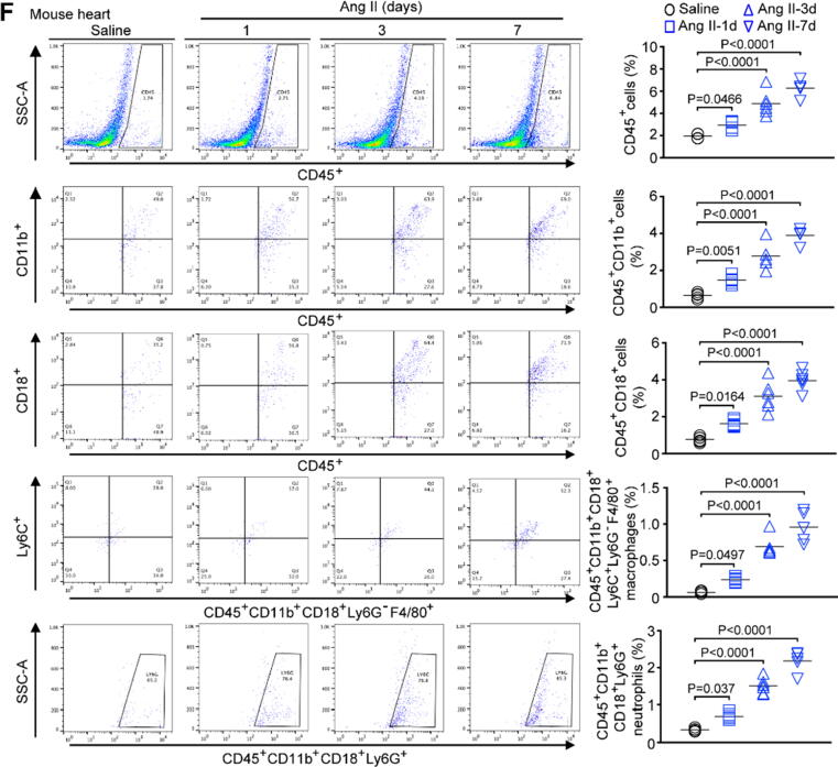
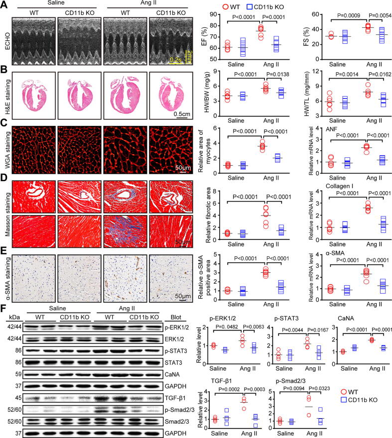
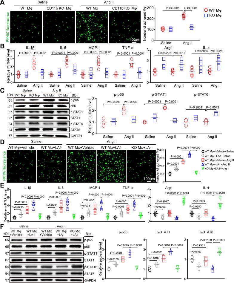
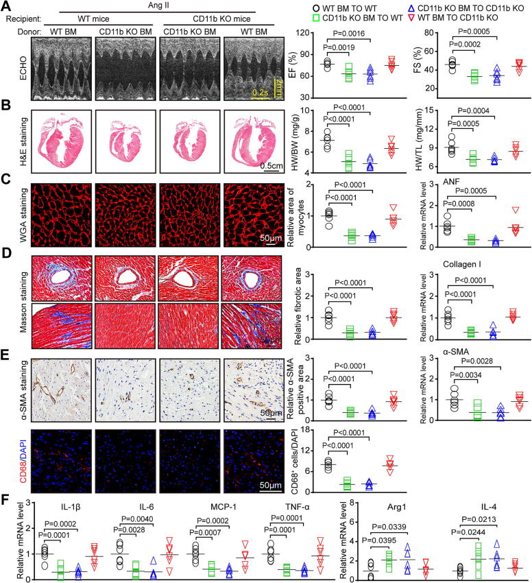
Results: Our microarray data showed that integrin subunits Itgam (CD11b) and Itgb2 (CD18) were the most highly upregulated in Ang II-infused hearts. CD11b expression and CD11b/CD18+ myelomonocytes were also time-dependently increased. KO or pharmacological blockade of CD11b greatly attenuated cardiac remodeling and macrophage infiltration and M1 polarization induced by Ang II or DOCA-salt. This protection was verified in wild-type mice transplanted with CD11b-deficient BM cells. Conversely, administration of CD11b agonist LA1 showed the opposite effects. Further, CD11b KO reduced Ang II-induced macrophage adhesion and M1 polarization, leading to reduction of cardiomyocyte enlargement and fibroblast differentiation in vitro. The numbers of CD14+CD11b+CD18+ monocytes and CD15+CD11b+CD18+ granulocytes were obviously higher in HF patients than in normal controls.

Conclusion: Our data demonstrate an important role of CD11b+ myeloid cells in hypertensive cardiac remodeling, and suggest that HF may benefit from targeting CD11b.

Keywords: Cardiac remodeling; Hypertensive stimuli; Integrin CD11b; Macrophage infiltration and polarization.


Conflict of interest statement

Declaration of Competing Interest The authors declare that they have no known competing financial interests or personal relationships that could have appeared to influence the work reported in this paper.

Figures

None
Graphical abstract
Fig. 1
Fig. 1
Ang II increased the level of CD11b/CD18 and its ligands, as well as CD11b/CD18+ myelomonocytes, in mouse hearts. A-B, WT mice were treated with Ang II for one, three and seven days. Heatmaps of cardiac α and β integrins and their ligands (n = 3). C, qPCR of CD11b, CD18, ICAM-1 and Factor X (n = 3). D, Western blot of CD11b and CD18 (top) and quantification (bottom, n = 4). E, Cardiac CD11b immunohistochemical staining (n = 6). F, Flow cytometric data of CD45+, CD45+CD11b+, CD45+CD18+, and CD45+CD11b+CD18+ myeloid cells, including Ly6C+Ly6G-F4/80+ MΦs and Ly6G+ neutrophils, in the aortas (left) and the percentage of each type of cell (n = 6). Data were presented as the mean ± SD and were analyzed with one-way ANOVA. Each P value is displayed in the image.
Fig. 1
Fig. 1
Ang II increased the level of CD11b/CD18 and its ligands, as well as CD11b/CD18+ myelomonocytes, in mouse hearts. A-B, WT mice were treated with Ang II for one, three and seven days. Heatmaps of cardiac α and β integrins and their ligands (n = 3). C, qPCR of CD11b, CD18, ICAM-1 and Factor X (n = 3). D, Western blot of CD11b and CD18 (top) and quantification (bottom, n = 4). E, Cardiac CD11b immunohistochemical staining (n = 6). F, Flow cytometric data of CD45+, CD45+CD11b+, CD45+CD18+, and CD45+CD11b+CD18+ myeloid cells, including Ly6C+Ly6G-F4/80+ MΦs and Ly6G+ neutrophils, in the aortas (left) and the percentage of each type of cell (n = 6). Data were presented as the mean ± SD and were analyzed with one-way ANOVA. Each P value is displayed in the image.
Fig. 2
Fig. 2
CD11b deficiency weakens Ang II-caused cardiac hypertrophy and fibrosis. WT and CD11b KO mice were treated with saline or Ang II for 14 days. A, Echocardiography (left) and EF% and FS% (right, n = 6). B, H&E staining (left) and quantification of the HW/BW and HW/TL ratios (right, n = 6). C, WGA staining (left) and quantification (right, n = 6); qPCR of ANF (n = 6). D, Masson's trichrome staining (left) and quantification (right, n = 6); qPCR of collagen I (n = 6). E, α-SMA immunohistochemical staining (left) and quantification (right, n = 6); qPCR of α-SMA (n = 6). F, Immunoblot of p-ERK1/2, ERK1/2, p-STAT3, STAT3, CaNA, TGF-β1, p-Smad2/3 and Smad2/3 (left) and quantification (right, n = 4). Data were expressed as the mean ± SD and were analyzed with two-way ANOVA. Each P value is displayed in the image.
Fig. 3
Fig. 3
CD11b KO ameliorates Ang II-induced CD11b-positive myelomonocyte infiltration and MΦ polarization. WT and CD11b KO mice were infused with saline or Ang II for 14 days. A, Flow cytometric data of CD45+ myelomonocytes, including CD11b+Ly6G-Ly6C+F4/80+ MΦs and CD11b+Ly6G+F4/80- neutrophils (left), in the heart, and quantification (right, n = 6). B, CD68 immunofluorescent staining (red) and quantification (right, n = 6). C, qPCR of IL-1β, IL-6, MCP-1, TNF-α, Arg1 and IL-4 (n = 6). D, Immunoblot of p-p65, p65, p-STAT1, STAT1, p-STAT6 and STAT6 (left) and quantification (right, n = 4). Data were expressed as the mean ± SD and were analyzed with two-way ANOVA. Each P value is displayed in the image. (For interpretation of the references to colour in this figure legend, the reader is referred to the web version of this article.)
Fig. 4
Fig. 4
CD11b deficiency inhibits Ang II-induced macrophage adhesion to ECs, as well as upregulation of M1 genes and downregulation of M2 genes in vitro, and CD11b activation with LA1 accelerates these effects. A, Macrophages were stained with PKH67 (green) and added to HUVECs that were prestimulated with Ang II for 24 h (left), and quantification of adherent cells (right, n = 3 independent experiments, 6 fields). B, qPCR of TNF-α, IL-1β, IL-6, MCP-1, Arg1 and IL-4 in macrophages (n = 3 independent experiments). C, Immunoblot of p65, STAT1 and STAT6 in macrophages (left) and quantification (right, n = 3 independent experiments). D, HUVECs were pretreated with Ang II for 24 h. Macrophages were stained with PKH67 (green), treated with the CD11b agonist LA1 for 30 min, and then added to the pretreated HUVECs. Representative images (left) and quantification of adherent cells on HUVECs (right, n = 3 independent experiments, 6 fields). E, qPCR of TNF-α, IL-1β, IL-6, MCP-1, Arg1 and IL-4 in macrophages (n = 3 independent experiments, 6 fields). F, Immunoblot of p65, STAT1 and STAT6 in macrophages (left) and quantification (right, n = 3 independent experiments). The data are expressed as m ± SD and were analyzed with two-way ANOVA followed by Sidak’s multiple comparison test. (For interpretation of the references to colour in this figure legend, the reader is referred to the web version of this article.)
Fig. 5
Fig. 5
BM-derived CD11b-deficient cells protect against Ang II-caused cardiac hypertrophy, fibrosis and inflammation. WT or CD11b KO mice were reconstituted with BM cells derived from WT or CD11b KO mice and treated with Ang II for 14 days. A, Echocardiography (left) and EF% and FS% (right, n = 6). B, H&E staining (left), and quantification of the HW/BW and HW/TL ratios (right, n = 6). C, WGA staining (left) and quantification (right, n = 6); qPCR of ANF (n = 6). D, Masson's trichrome staining (left) and quantification (right, n = 6); qPCR of collagen I (n = 6). E, α-SMA immunohistochemical staining (left) and quantification (right, n = 6); qPCR of α-SMA (n = 6); CD68 immunofluorescent staining (red) and quantification (right, n = 6). F, qPCR of IL-1β, IL-6, MCP-1, TNF-α, Arg1 and IL-4 (n = 6). Data were expressed as the mean ± SD and were analyzed with two-way ANOVA. Each P value is displayed in the image. (For interpretation of the references to colour in this figure legend, the reader is referred to the web version of this article.)
Fig. 6
Fig. 6
Pharmacological blockade of CD11b prevents Ang II-caused cardiac remodeling. WT mice were administered CD11b mAb or IgG control and treated with Ang II for 14 days. A, H&E staining (left) and quantification of HW/BW and HW/TL ratios (right, n = 6). B, WGA staining (left) and quantitation (right, n = 6); qPCR of ANF (n = 6). C, Masson's trichrome staining (left) and quantification (right, n = 6); qPCR of collagen I (n = 6). D, α-SMA immunohistochemical staining (left) and quantification (right, n = 6); qPCR of α-SMA (n = 6); CD68 immunofluorescent staining (red) and quantification (right, n = 6). E, qPCR of IL-1β, IL-6, MCP-1, TNF-α, Arg1 and IL-4 (n = 6). F, Echocardiography (left) and EF% and FS% (right, n = 6). Data were expressed as the mean ± SD and were analyzed with two-way ANOVA. Each P value is displayed in the image. (For interpretation of the references to colour in this figure legend, the reader is referred to the web version of this article.)
Fig. 7
Fig. 7
CD11b/CD18 positive myelomonocytes and the levels of ligands are increased in the blood of patients with HF. A, Flow cytometric analysis of CD45+ myelomonocytes, including CD11b+, CD18+, and CD11b+CD18+ cells; CD14+CD11b+, CD14+CD18+ and CD14+CD11b+CD18+ monocytes; and CD15+CD11b+, CD15+CD18+ and CD15+CD11b+CD18+ granulocytes, in the blood of HF subjects (n = 65) and controls (n = 65). B, ELISA of blood ICAM-1, fibrinogen a and factor X levels in the blood of HF subjects (n = 65) and controls (n = 65). Data were expressed as the mean ± SD and were analyzed with a sample t test. Each P value is displayed in the image.
Fig. 8
Fig. 8
A graphical abstract for CD11b contributes to cardiac hypertensive remodeling. Ang II stimulates the endothelial cells to release abundant CAMs, including ICAM-1, factor X and fibrinogen a, which recruit bone marrow-derived CD11b/CD18+ monocytes. These cells migrate and adhere to the vascular endothelium, then infiltrate into heart and differentiate into CD11b/CD18+ M1 macrophages that produce large amount of proinflammatory cytokines (IL-1β, IL-6 and TNF-α), thereby promoting cardiac remodeling and dysfunction. Ablation or inhibition of CD11b with neutralizing antibody significantly attenuates these effects.

References

    1. González A., Ravassa S., López B., Moreno M.U., Beaumont J., San José G. Myocardial Remodeling in Hypertension. Hypertension. 2018;72(3):549–558. - PubMed
    1. Nwabuo C.C., Vasan R.S. Pathophysiology of Hypertensive Heart Disease: Beyond Left Ventricular Hypertrophy. Curr Hypertens Rep. 2020;22(2):11. - PubMed
    1. Mouton A.J., Li X., Hall M.E., Hall J.E. Obesity, Hypertension, and Cardiac Dysfunction: Novel Roles of Immunometabolism in Macrophage Activation and Inflammation. Circ Res. 2020;126(6):789–806. - PMC - PubMed
    1. Harjunpää H., Llort Asens M., Guenther C., Fagerholm S.C. Cell Adhesion Molecules and Their Roles and Regulation in the Immune and Tumor Microenvironment. Front Immunol. 2019;10:1078. - PMC - PubMed
    1. Hu C., Zhang X., Zhang N., Wei W.Y., Li L.L., Ma Z.G., et al. Osteocrin attenuates inflammation, oxidative stress, apoptosis, and cardiac dysfunction in doxorubicin-induced cardiotoxicity. Clin Transl Med. 2020;10(3):e124. - PMC - PubMed

Publication types

LinkOut - more resources