. 2023 Apr 1;1864(2):148962.
doi: 10.1016/j.bbabio.2023.148962. Epub 2023 Feb 21.

Mutational analysis of a conserved positive charge in the c-ring of E. coli ATP synthase

Affiliations

Mutational analysis of a conserved positive charge in the c-ring of E. coli ATP synthase

Rashmi K Shrestha et al. Biochim Biophys Acta Bioenerg. .

Abstract

F1Fo ATP synthase is a ubiquitous molecular motor that utilizes a rotary mechanism to synthesize adenosine triphosphate (ATP), the fundamental energy currency of life. The membrane-embedded Fo motor converts the electrochemical gradient of protons into rotation, which is then used to drive the conformational changes in the soluble F1 motor that catalyze ATP synthesis. In E. coli, the Fo motor is composed of a c10 ring (rotor) alongside subunit a (stator), which together provide two aqueous half channels that facilitate proton translocation. Previous work has suggested that Arg50 and Thr51 on the cytoplasmic side of each subunit c are involved in the proton translocation process, and positive charge is conserved in this region of subunit c. To further investigate the role of these residues and the chemical requirements for activity at these positions, we generated 13 substitution mutants and assayed their in vitro ATP synthesis, H+ pumping, and passive H+ permeability activities, as well as the ability of mutants to carry out oxidative phosphorylation in vivo. While polar and hydrophobic mutations were generally tolerated in either position, introduction of negative charge or removal of polarity caused a substantial defect. We discuss the possible effects of altered electrostatics on the interaction between the rotor and stator, water structure in the aqueous channel, and interaction of the rotor with cardiolipin.

Keywords: ATP synthase; Cardiolipin; Oxidative phosphorylation; Proton transport; Site-directed mutagenesis.


Conflict of interest statement

Declaration of competing interest The authors declare that they have no known competing financial interests or personal relationships that could have appeared to influence the work reported in this paper.

Figures

Figure 1:
Figure 1:. Structural context of Arg50 and Thr51.
Structure of E. coli ATP synthase (PDB ID: 6OQW) with detail showing the a-c interface of Fo (PDB ID: 6VWK) as viewed from the cytoplasmic (N) side. Approximate side chain positions are shown for cArg50 and cThr51, as well as for the essential aArg210 and cAsp61 residues for context. The approximate extent of the N-side proton channel is shown in light blue.
Figure 2:
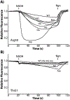
Figure 2:. H+ pumping and ATP synthesis activities of Arg50 mutants.
A) Representative ACMA fluorescence time traces show ATP-driven H+ pumping activity. Markers indicate the addition of ATP at 20s and the addition of nigericin (Ngn) at 100s. Fluorescence values for each trace were normalized to the initial value. B) Representative luminescence time traces are shown for H+ gradient-driven ATP synthesis activity. Inverted membrane vesicles were energized with NADH at t=0, and luminescence (arbitrary units) was monitored for 10 min. C) Activities of Arg50 mutants are plotted relative to WT. Pumping activity (dark bars) is the percent fluorescence quenching (see Section 4.3). ATP synthesis activity (light bars) is the maximum slope (luminescence/min) as defined by five consecutive points (about 3 min). Activities are plotted as mean ± standard error (n≥3).
Figure 3:
Figure 3:. H+ pumping and ATP synthesis activities of Thr51 mutants.
A) Representative ACMA fluorescence time traces. Markers indicate the addition of ATP at 20s and the addition of nigericin (Ngn) at 100s. Fluorescence values for each trace were normalized to the initial value. B) Representative luminescence time traces are shown for H+ gradient-driven ATP synthesis. Inverted membrane vesicles were energized with NADH at t=0, and luminescence (arbitrary units) was monitored in real time for 10 min. C) Activities of Thr51 mutants are plotted relative to WT. Pumping activity (dark bars) is the percent fluorescence quenching (see Section 4.3). ATP synthesis activity (light bars) is the maximum slope (luminescence/min) defined by five consecutive points (about 3 min). Activities are plotted as mean ± standard error (n≥3).
Figure 4:
Figure 4:. H+ pumping and ATP synthesis activities of the Arg50Ala/Thr51Ala mutant.
A) Representative ACMA fluorescence time traces. Markers indicate the addition of ATP at 20s and the addition of nigericin (Ngn) at 100s. Fluorescence values for each trace were normalized to the initial value. B) Representative luminescence time traces are shown for H+ gradient-driven ATP synthesis. Inverted membrane vesicles were energized with NADH at t=0, and luminescence (arbitrary units) was monitored in real time for 10 min. C) Activities of Thr51 mutants are plotted relative to WT. Pumping activity (dark bars) is the percent fluorescence quenching (see Section 4.3). ATP synthesis activity (light bars) is the maximum slope (luminescence/min) defined by five consecutive points (about 3 min). Activities are plotted as mean ± standard error (n≥3).
Figure 5:
Figure 5:. Effects of substitutions on passive H+ translocation.
Representative ACMA fluorescence time traces are shown for stripped membrane vesicles of Arg50 substitutions (A) and Thr51 substitutions (B). Markers indicate the addition of NADH at 20 s and the addition of nigericin (Ngn) at 100 s. Fluorescence values for each trace were normalized to the initial value.
Figure 6:
Figure 6:. Effects of mutations on succinate growth.
E. coli mutants, including wild type (WT) and DK8 (Δunc) were grown on M63-TIV minimal medium supplemented with 0.6% succinate (see Section 4.6) and cell density was monitored by OD550 over 8 hours. Maximum cell density during the growth period was normalized to WT. Bars represent mean ± standard error (n≥3) relative to WT.
Figure 7:
Figure 7:. No effect of mutations on insertion of Fo subunits.
Western blots of WT and mutant inverted membrane vesicles using antisera against subunit a. DK8 (Δunc) and Gly23Asp were included as negative controls. Uncropped blots, including molecular weight standards, are shown in Figure S2.
Figure 8:
Figure 8:. Conservation and possible interactions of Arg50 and Thr51.
A) Amino acid sequence alignment of subunit c from several species in the region between the conserved R(Q/N)P loop motif and H+ binding acidic residue (open boxes). Arg and Lys residues in this region are highlighted in blue and nearby Ser and Thr residues are highlighted in green. EC, Escherichia coli; BS, Bacillus subtilis; PM, Propionigenium modestum; MT, Mycobacterium tuberculosis; SC, Saccharomyces cerevisiae; HS, Homo sapiens; SO, Spinacia oleracea; AT, Arabidopsis thaliana. B) Structural detail of the a-c interface (PDB ID: 6VWK) showing proximity of Arg50 and Thr51 to negative charges in subunit a, including Asp92 and the acidic C-terminal residues. Note that side chain positions are approximate, and the highlighted C-terminal residue (ball and stick representation) is Glu269, since Glu270 and His271 are not included in the model. C) Approximate placement of a cardiolipin headgroup next to a single c subunit (PDB ID: 6VWK) showing potential interactions consistent with the cardiolipin binding site proposed by Corey et al. [31].

References

    1. Kühlbrandt W, Structure and Mechanisms of F-Type ATP Synthases, Annu. Rev. Biochem, 88 (2019) 515–549. - PubMed
    1. Giorgio V, von Stockum S, Antoniel M, Fabbro A, Fogolari F, Forte M, Glick GD, Petronilli V, Zoratti M, Szabó I, Lippe G, Bernardi P, Dimers of mitochondrial ATP synthase form the permeability transition pore, Proc. Natl. Acad. Sci, 110 (2013) 5887–5892. - PMC - PubMed
    1. Carraro M, Checchetto V, Szabó I, Bernardi P, F- ATP synthase and the permeability transition pore: fewer doubts, more certainties, FEBS Lett, 593 (2019) 1542–1553. - PubMed
    1. Nesci S, Trombetti F, Algieri C, Pagliarani A, A Therapeutic Role for the F1FO-ATP Synthase, SLAS Discov., 24 (2019) 893–903. - PubMed
    1. Hards K, Cook GM, Targeting bacterial energetics to produce new antimicrobials, Drug Resist. Updat, 36 (2018) 1–12. - PubMed

Publication types

LinkOut - more resources