

Medullary Tegmental Cap Dysplasia: Fetal and Postnatal Presentations of a Unique Brainstem Malformation
- PMID: 36822823
- PMCID: PMC10187821
- DOI: 10.3174/ajnr.A7805
Medullary Tegmental Cap Dysplasia: Fetal and Postnatal Presentations of a Unique Brainstem Malformation
Abstract
Background and purpose: Medullary tegmental cap dysplasia is a rare brainstem malformation, first described and defined by James Barkovich in his book Pediatric Neuroimaging from 2005 as an anomalous mass protruding from the posterior medullary surface. We describe the neuroimaging, clinical, postmortem, and genetic findings defining this unique malformation.
Materials and methods: This is a multicenter, international, retrospective study. We assessed the patients' medical records, prenatal ultrasounds, MR images, genetic findings, and postmortem results. We reviewed the medical literature for all studies depicting medullary malformations and evaluated cases in which a dorsal medullary protuberance was described.
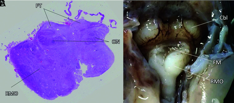
Results: We collected 13 patients: 3 fetuses and 10 children. The medullary caps had multiple characteristics. Associated brain findings were a rotated position of the medulla, a small and flat pons, cerebellar anomalies, a molar tooth sign, and agenesis of the corpus callosum. Systemic findings included the following: polydactyly, hallux valgus, large ears, and coarse facies. Postmortem analysis in 3 patients revealed that the cap contained either neurons or white matter tracts. We found 8 publications describing a dorsal medullary protuberance in 27 patients. The syndromic diagnosis was Joubert-Boltshauser syndrome in 11 and fibrodysplasia ossificans progressiva in 14 patients.
Conclusions: This is the first study to describe a series of 13 patients with medullary tegmental cap dysplasia. The cap has different shapes: distinct in Joubert-Boltshauser syndrome and fibrodysplasia ossificans progressive. Due to the variations in the clinical, imaging, and postmortem findings, we conclude that there are multiple etiologies and pathophysiology. We suggest that in some patients, the pathophysiology might be abnormal axonal guidance.
© 2023 by American Journal of Neuroradiology.
Figures


Similar articles
-
Isolated Unilateral Cerebellar Hemispheric Dysplasia: A Rare Entity.Can J Neurol Sci. 2019 Nov;46(6):760-761. doi: 10.1017/cjn.2019.249. Can J Neurol Sci. 2019. PMID: 31352912
-
Prenatal diagnosis of rhombencephalosynapsis: neuroimaging features and severity of vermian anomaly.Ultrasound Obstet Gynecol. 2021 Dec;58(6):864-874. doi: 10.1002/uog.23660. Ultrasound Obstet Gynecol. 2021. PMID: 33942916
-
Pontine tegmental cap dysplasia: the role of diffusion tensor imaging.BMJ Case Rep. 2023 Nov 22;16(11):e253556. doi: 10.1136/bcr-2022-253556. BMJ Case Rep. 2023. PMID: 37993144
-
Pontine Tegmental Cap Dysplasia in an Extremely Preterm Infant and Review of the Literature.J Child Neurol. 2017 Mar;32(3):334-340. doi: 10.1177/0883073816680748. Epub 2016 Dec 20. J Child Neurol. 2017. PMID: 28193110 Review.
-
Role of magnetic resonance imaging in fetuses with mild or moderate ventriculomegaly in the era of fetal neurosonography: systematic review and meta-analysis.Ultrasound Obstet Gynecol. 2019 Aug;54(2):164-171. doi: 10.1002/uog.20197. Epub 2019 Jul 11. Ultrasound Obstet Gynecol. 2019. PMID: 30549340
Cited by
-
Prenatal assessment of brain malformations on neuroimaging: an expert panel review.Brain. 2024 Dec 3;147(12):3982-4002. doi: 10.1093/brain/awae253. Brain. 2024. PMID: 39054600 Free PMC article. Review.
-
Dandy-Walker Phenotype with Brainstem Involvement: 2 Distinct Subgroups with Different Prognosis.AJNR Am J Neuroradiol. 2023 Oct;44(10):1201-1207. doi: 10.3174/ajnr.A7967. Epub 2023 Aug 17. AJNR Am J Neuroradiol. 2023. PMID: 37591769 Free PMC article.
References
-
- Barkovich AJ. Pediatric Neuroimaging. 5th ed. Lippincott Williams & Wilkins; 2005:932
Publication types
MeSH terms
Supplementary concepts
LinkOut - more resources
Full Text Sources
Medical
Miscellaneous