. 2023 Feb 23;13(1):3166.
doi: 10.1038/s41598-023-30240-8.

Antibacterial and antioxidant double-layered nanofibrous mat promotes wound healing in diabetic rats

Affiliations

Antibacterial and antioxidant double-layered nanofibrous mat promotes wound healing in diabetic rats

Fereshteh Nejaddehbashi et al. Sci Rep. .

Abstract

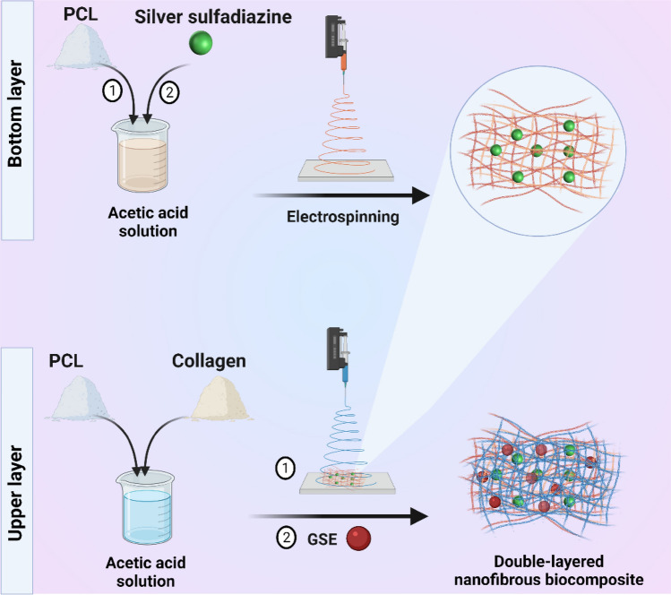
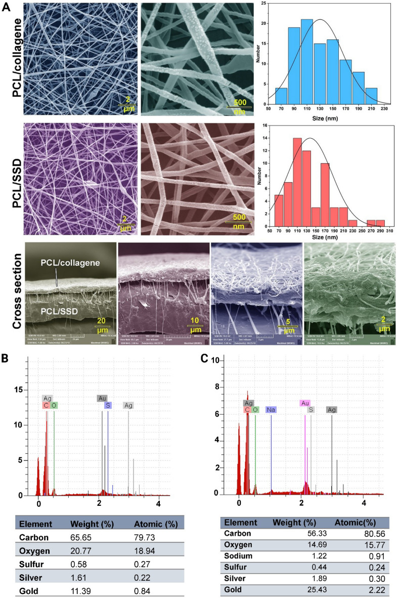
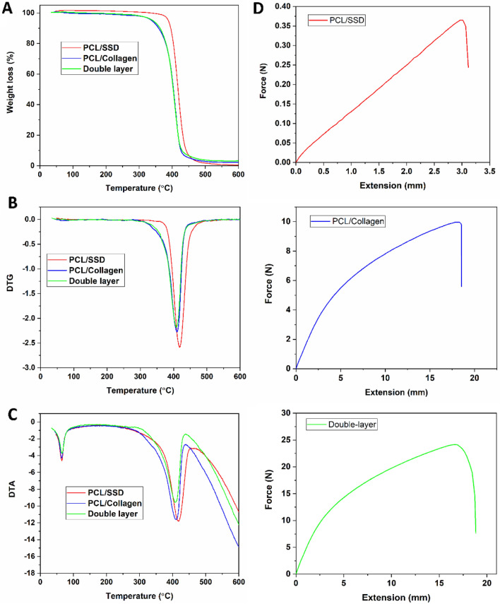
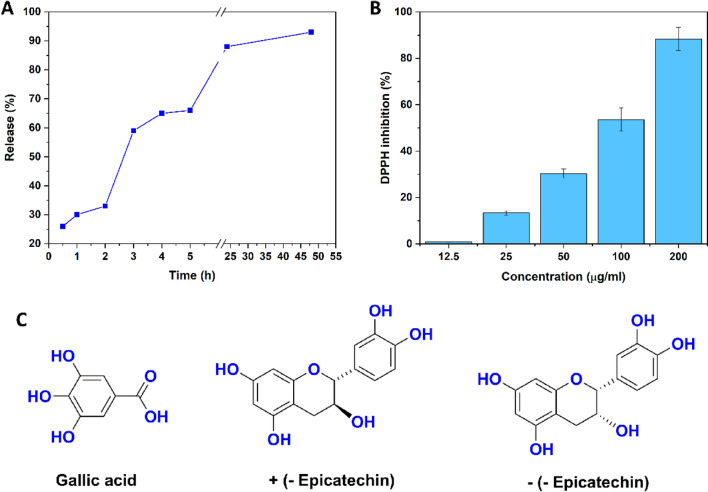
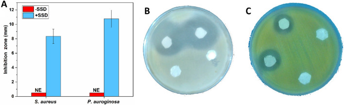
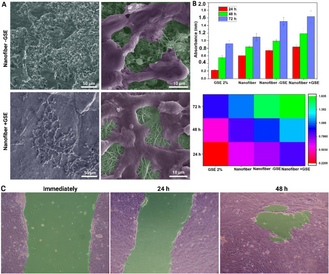
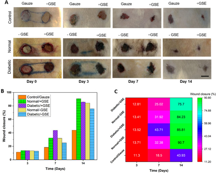
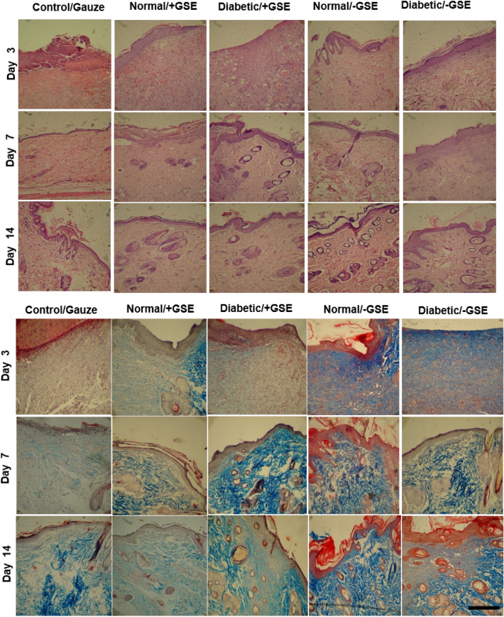
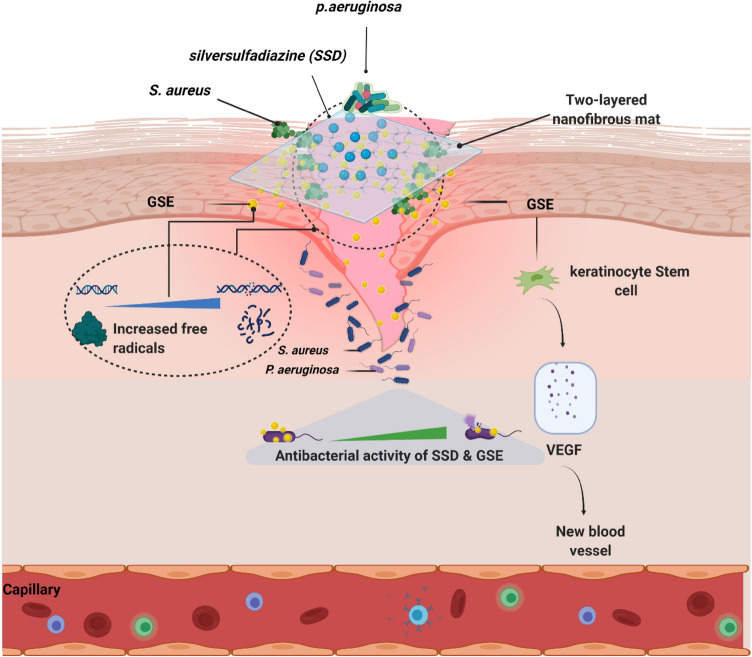
Diabetic wounds are problematic to heal owing to microbial infections as well as decreased proliferation and high concentrations of reactive oxygen species. In this study, a double-layered nanofibrous mat containing grape seed extract (GSE) and silver sulfadiazine (SSD) was fabricated. A synthetic biodegradable polymer, e.g., polycaprolactone (PCL), and a natural material (i.e., collagen) were employed as wound dressing substances. The results showed that GSE possesses antioxidant activity which can be helpful in reducing free radicals. The platform exhibited antibacterial activity against gram-positive and -negative bacteria. The double-layered nanofibrous mat containing GSE and SSD not only was not toxic but also amplified the cell proliferation compared to a pure mat, showing the effect of plant extract. After induction of a round wound, the animals were divided into three groups, namely (1) normal group (receiving + GSE/-GSE nanofiber), (2) diabetic group (receiving + GSE/-GSE nanofiber), and (3) control group (receiving gauze). In vivo evaluation demonstrated no significant differences in the healing process of normal rats. Surprisingly, fully repaired skin was observed on day 14 in the double-layered nanofibrous mat containing GSE in the normal and diabetic groups whereas the wound of diabetic rats treated with pure mat was not completely healed. The macroscopic and microscopic results after 14 days showed the following order in wound repair: Normal/ + GES > Diabetic/ + GSE > Normal/-GES > Diabetic/-GSE > control (with gauze) (p < 0.05). Accordingly, the double-layered nanofibrous mat containing GSE and SSD used in the present study could be considered as a suitable wound dressing in order to shorten healing time and prevent infection during the wound healing process.


Conflict of interest statement

The authors declare no competing interests.

Figures

Figure 1
Figure 1
Manufacturing process of the scaffold via electrospining. Schematic illustration of fabrication of the double-layered nanofibrous mat.
Figure 2
Figure 2
Morphological characteristics of the double-layered nanofibrous mat with FE-SEM. (A) the top layer is PCL/Coll and the bottom layer is PCL/SSD, which are shown with size distribution. The placement of the double-layered nanofibrous mat is shown in the bottom-most panel. The presence of nanofiber filaments in the cross-section between the two layers indicates that the two layers are connected to each other. Elemental analysis was done on the double-layered nanofibrous mat without GSE (B) and the double-layered nanofibrous mat with GSE (C).
Figure 3
Figure 3
Thermal and mechanical properties of the electrospun nanofibers. (A) TGA, (B) DTG, and (C) DTA graphs of the samples. (D) Tensile test of the electrospun nanofibers.
Figure 4
Figure 4
Release profile and antioxidant activity. (A) Release profile of the GSE from the nanofibrous mat. (B) Antioxidant activity of GSE. (C) Some available compounds responsible for antioxidant activity of the GSE.
Figure 5
Figure 5
The antibacterial effect of the prepared nanofibrous mats against S. aureus and P. aeruginosa is shown in a bar graph (A). The inhibition zone against the nanofibrous mat with and without SSD in S. aureus (B) and P. aeruginosa is presented (C).
Figure 6
Figure 6
(A) FE-SEM image of HDF stem cells proliferation on the nanofibrous mat with and without GSE. The cells were artificially colored to be more distinguishable in the image. (B) Bar graph and heatmap related to the results of the MTT assay. (C) In vitro cell migration assay at different times. The images were colored to show the different areas.
Figure 7
Figure 7
Macroscopic evaluation of wound healing in normal and diabetic rats. (A) Models on days 0, 3, 7, and 14 following surgery, demonstrating the wound's gradual healing over time. Scale bar represents 10 mm. (B) Wound closure percentage and heatmap graph in diabetic and normal groups with and without extract during days 3, 7 and 14 of the repair processes. (C) GSE represents treatment without grape seed extract; + GSE represents treatment with seed extract. Scale bar represents 10 mm.
Figure 8
Figure 8
Microscopic evaluation of wound healing. Hematoxylin–eosin staining (A) and trichromassonʼs staining (B). Scale bar represents 200 µm.
Figure 9
Figure 9
Schematic illustration of the application of a double-layered nanofibrous mat with antibacterial and antioxidant properties to promote wound healing.

References

    1. Jamaledin R, Yiu CKY, Zare EN, Niu LN, Vecchione R, Chen G, Gu Z, Tay FR, Makvandi P. Advances in antimicrobial microneedle patches for combating infections. Adv. Mater. 2020;32(33):2002129. doi: 10.1002/adma.202002129. - DOI - PubMed
    1. Bagheri M, Validi M, Gholipour A, Makvandi P, Sharifi E. Chitosan nanofiber biocomposites for potential wound healing applications: antioxidant activity with synergic antibacterial effect. Bioeng. Transl. Med. 2022;7:e10254. doi: 10.1002/btm2.10254. - DOI - PMC - PubMed
    1. Makvandi P, Jamaledin R, Chen G, Baghbantaraghdari Z, Zare EN, Di Natale C, Onesto V, Vecchione R, Lee J, Tay FR. Stimuli-responsive transdermal microneedle patches. Mater. Today. 2021;47:206–222. doi: 10.1016/j.mattod.2021.03.012. - DOI - PMC - PubMed
    1. Makvandi P, Jamaledin R, Chen G, Baghbantaraghdari Z, Zare EN, Di Natale C, Onesto V, Vecchione R, Lee J, Tay FR. Stimuliponsive transdermal microneedle patches-res. Mater. Today. 2021;47:206–222. doi: 10.1016/j.mattod.2021.03.012. - DOI - PMC - PubMed
    1. Burford, G., Bodeker, G., Ryan, T.J. Skin and wound care: traditional, complementary and alternative medicine in public health dermatology. In: Traditional, Complementary And Alternative Medicine: Policy and Public Health Perspectives. pp. 311–348. World Scientific (2007)

Publication types

MeSH terms