. 2023 Apr;20(4):404-418.
doi: 10.1038/s41423-023-00982-6. Epub 2023 Feb 24.

HTR2A agonists play a therapeutic role by restricting ILC2 activation in papain-induced lung inflammation

Affiliations

HTR2A agonists play a therapeutic role by restricting ILC2 activation in papain-induced lung inflammation

Zhishuo Wang et al. Cell Mol Immunol. 2023 Apr.

Abstract

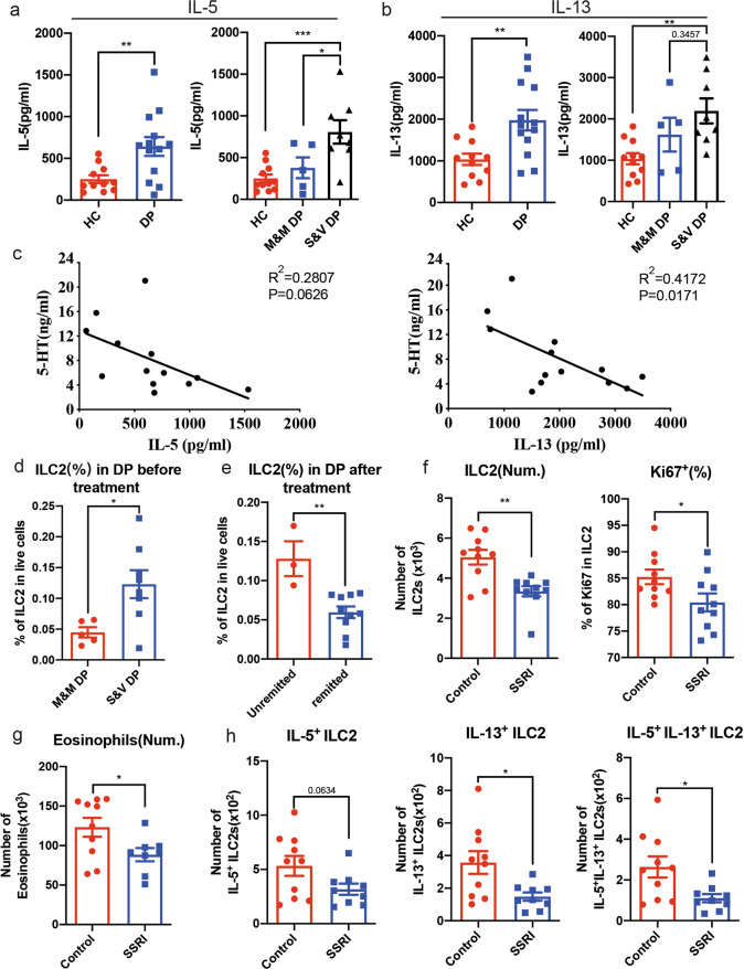
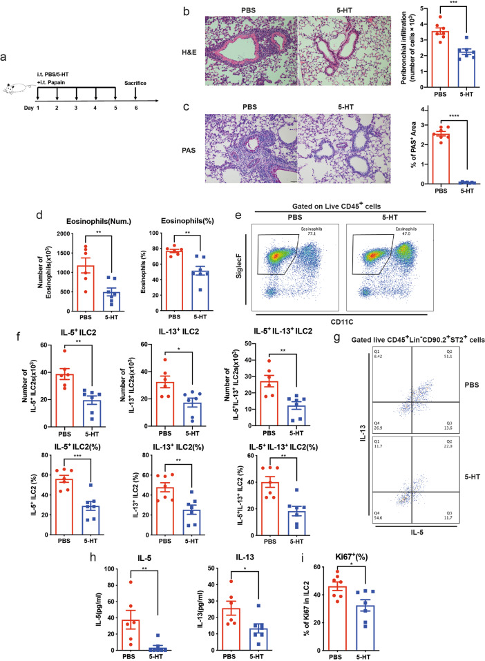
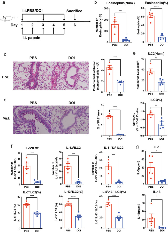
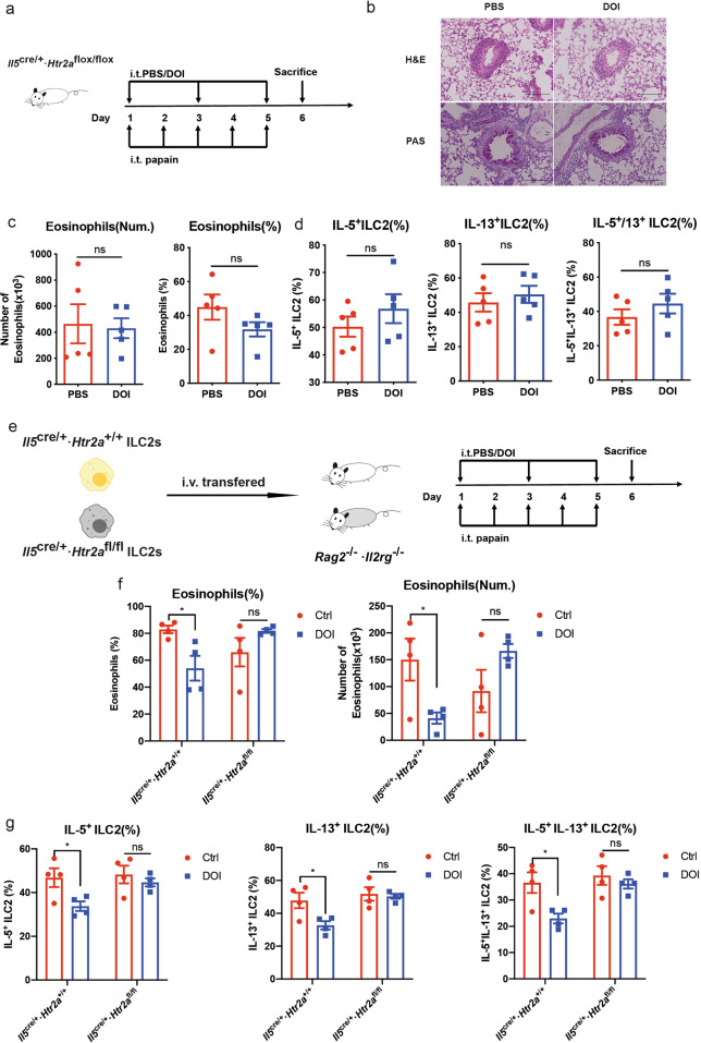
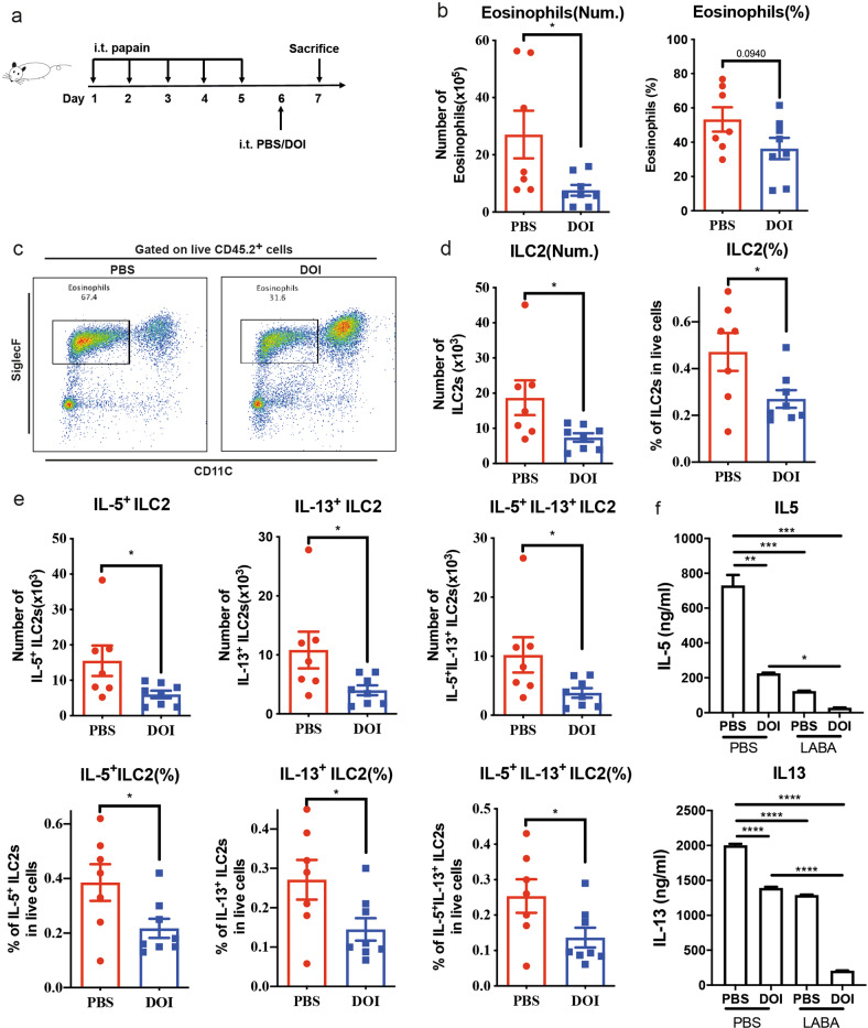
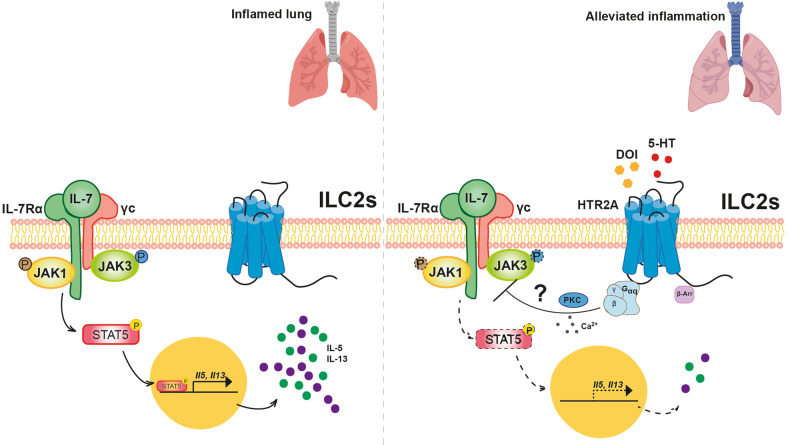
Group 2 innate lymphoid cells (ILC2s) are a category of heterogeneous cells that produce the cytokines IL-5 and IL-13, which mediate the type 2 immune response. However, specific drug targets on lung ILC2s have rarely been reported. Previous studies have shown that type 2 cytokines, such as IL-5 and IL-13, are related to depression. Here, we demonstrated the negative correlation between the depression-associated monoamine neurotransmitter serotonin and secretion of the cytokines IL-5 and IL-13 by ILC2s in individuals with depression. Interestingly, serotonin ameliorates papain-induced lung inflammation by suppressing ILC2 activation. Our data showed that the serotonin receptor HTR2A was highly expressed on ILC2s from mouse lungs and human PBMCs. Furthermore, an HTR2A selective agonist (DOI) impaired ILC2 activation and alleviated the type 2 immune response in vivo and in vitro. Mice with ILC2-specific depletion of HTR2A (Il5cre/+·Htr2aflox/flox mice) abolished the DOI-mediated inhibition of ILC2s in a papain-induced mouse model of inflammation. In conclusion, serotonin and DOI could restrict the type 2 lung immune response, indicating a potential treatment strategy for type 2 lung inflammation by targeting HTR2A on ST2+ ILC2s.

Keywords: DOI; Group 2 innate lymphoid cell; HTR2A; Serotonin (5-HT); Type 2 lung inflammation.


Conflict of interest statement

The authors declare no competing interests.

Figures

Fig. 1
Fig. 1
IL-33 mediated ILC2 activation in PBMCs from depressive patients. The type 2 immune response secretion of the cytokines a IL-5 and b IL-13 from PBMCs after IL-33 activation. c Analysis of the correlations between IL-5/IL-13 and 5-HT in the plasma of depressive patients. PBMCs were isolated from healthy control subjects and depressive patients and were then cultured in vitro with hIL-2 (100 U/ml), hIL-7 (20 ng/ml) and hIL-33 (50 ng/ml). The supernatants were collected, and the levels of the cytokines IL-5 and IL-13 were detected by ELISA. PBMCs with IL-5 secretion after IL-2/IL-7/IL-33 treatment that was one-fold higher than that after IL-2/IL-7 treatment were used for data analysis. Patients were separated into the severe/very severe depression patient (S&V DP) group and the mild/moderate depression patient (M&M DP) group according to the Hamilton Rating Scale for Depression (HAMD-17) total score. d The percentage of ILC2s in the PBMCs from depressive patients before treatment. e The percentage of ILC2s in the PBMCs from unremitted and remitted depressive patients. f The number and proliferation of lung ILC2s in the papain-induced type 2 immune response model after SSRI treatment. g The number of lung eosinophils in the papain-induced type 2 immune response model after SSRI treatment. h Flow cytometric analysis of the numbers of IL-5+, IL-13+ and IL-5+IL-13+ cells among lung ILC2s after stimulation with PMA, ionomycin and BFA for 4 h. The mice were intratracheally challenged with papain from Day 6 to Day 10 and treated with the SSRI escitalopram in 0.9% NaCl solution (i.p. 10 mg/kg) or 0.9% NaCl solution only from Day 1 to Day 10. The bars and error bars show the means ± SEMs; *P < 0.05, **P < 0.01, ***P < 0.001 by unpaired Student’s t test (a, b, d, e, f, g, h) or one-way ANOVA with Tukey’s multiple comparisons test (a, b). Data are representative of three independent experiments
Fig. 2
Fig. 2
Serotonin inhibited ILC2 function in a papain-induced mouse model of inflammation in vivo. a Mice were intratracheally challenged with papain together with PBS or serotonin (10 μg/kg) for 5 consecutive days and sacrificed on Day 6. b Representative H&E-stained images of lung sections (bars, 50 μm) (left) and histological quantification of peribronchial inflammatory cell infiltration by ImageJ (right). c Representative PAS staining of lung sections (bars, 50 μm) (left) and histological quantification of the PAS+ (positive) area by ImageJ (right). d Quantification and e representative flow cytometry analysis of the total eosinophils (live CD45+CD11c−/loSiglecF+) in BALF (n = 6 or 7 per group). f Quantification and g representative flow cytometry analysis of IL-5 and IL-13 production in lung ILC2s stimulated with PMA plus ionomycin and BFA for 4 h. h The levels of IL-5 and IL-13 in BALF were measured by ELISA. i Ki67 analysis of lung LinCD90.2+ST2+ ILC2 cells by flow cytometry. The bars and error bars show the means ± SEMs; *P < 0.05; **P < 0.01; ***P < 0.001; ****P < 0.0001 by unpaired Student’s t test. Data are representative of three independent experiments
Fig. 3
Fig. 3
Lung ST2+ ILC2s highly specifically express Htr2a, and DOI, a selective 5-HTR2A agonist, can also inhibit ILC2 activation in vitro. a Relative expression of 5-Htrs in mouse lung ILC2s. b Htr2a expression in different cell types sorted from naïve mice. Lung ILC2s were sorted as live CD45+LinCD127+ST2+ cells. c Relative expression of Htr2a in different cell types sorted from human PBMCs. d Quantification and e representative flow cytometry analysis of IL-5+, IL-13+ and IL-5+/13+ ILC2s upon treatment with different dosages of DOI. f The amounts of IL-5 and IL-13 secreted from ILC2s into the culture medium measured by ELISA. g Flow cytometric analysis of ILC2 viability with FVS780 staining with or without DOI treatment. h Flow cytometric analysis of the percentages of live and apoptotic ILC2s treated with or without DOI with PI/Annexin V staining. i Flow cytometry analysis of ILC2 cell viability after treatment with or without DOI by CCK-8 cytotoxicity assay. The bars and error bars show the means ± SEMs; *P < 0.05; **P < 0.01; ***P < 0.001; ****P < 0.0001; ns, not significant by ordinary one-way ANOVA with multiple comparisons (d, f, g) or unpaired Student’s t test (h, i). Data are representative of three independent experiments
Fig. 4
Fig. 4
DOI can control ILC2-dominated lung inflammation through the highly expressed 5-HT receptor 2A on lung ST2+ ILC2s in vivo. a Schematic of DOI administration to papain-treated wild-type mice. Mice were intratracheally challenged with DOI or PBS every two days followed by papain administration for 5 consecutive days and then sacrifice on Day 6. b Quantification of total eosinophils (live CD45+CD11c−/loSiglecF+) and their percentages in BALF (n = 5 per group). c Representative H&E-stained images of lung sections (bars, 100 μm) (left) and histological quantification of peribronchial inflammatory cell infiltration by ImageJ (right). d Representative PAS staining of lung sections (bars, 100 μm) (left) and histological quantification of PAS+ (positive) area by ImageJ (right). e Quantification of the flow cytometric analysis of lung LinCD90.2+ST2+ ILC2 cell numbers and percentages in this model. f Flow cytometric analysis of IL-5 and IL-13 in lung ILC2s stimulated with PMA plus ionomycin and BFA for 4 h. g The levels of IL-5 and IL-13 in BALF were measured by ELISA. The bars and error bars show the means ± SEMs; *P < 0.05; **P < 0.01; ***P < 0.001; ****P < 0.0001 by unpaired Student’s t test. Data are representative of three independent experiments
Fig. 5
Fig. 5
HTR2A, rather than other serotonin receptors, mediated the inhibition of lung ST2+ ILC2s. a Schematic of DOI administration to papain-induced Il5cre/+·Htr2aflox/flox mice. Mice were intratracheally challenged with DOI or PBS every two days followed by papain administration for 5 consecutive days and then sacrifice on Day 6. b Representative H&E and PAS staining images of lung sections (bars, 100 μm). c Quantification of total eosinophils (live CD45+CD11c−/loSiglecF+) and their percentages in BALF (n = 5 per group). d Flow cytometric analysis of IL-5 and IL-13 in lung ILC2s stimulated with PMA plus ionomycin and BFA for 4 h. e Schematic of DOI administration to papain-induced Rag2−/−·Il2rg−/− mice receiving Il5cre/+·Htr2a+/+ and Il5cre/+·Htr2aflox/flox ILC2s (i.v. 5 × 105 ILC2s/mouse). f The percentages and numbers of eosinophils in the BALF of Rag2−/−·Il2rg−/− mice receiving Il5cre/+·Htr2a+/+ and Il5cre/+·Htr2aflox/flox ILC2s treated with or without DOI in the papain-induced lung inflammation model (n = 4 per group). g Flow cytometric analysis of the levels of IL-5 and IL-13 in lung ILC2s stimulated with PMA plus ionomycin and BFA for 4 h. The bars and error bars show the means ± SEMs. *P < 0.05; ns, not significant by unpaired Student’s t test (b, c) or two-way ANOVA (f, g)
Fig. 6
Fig. 6
DOI could be a therapeutic drug for ILC2-mediated type 2 lung inflammation. a Schematic of therapeutic DOI administration after papain-induced lung inflammation. Mice were intratracheally challenged with a single dose of DOI or PBS on Day 6 after 5 consecutive days of papain treatment, and mice were sacrificed on Day 7. b Quantification of total eosinophils (live CD45+CD11c−/loSiglecF+) and their percentages in BALF (n = 7 or 8 per group). c Representative flow cytometry analysis of total eosinophils (live CD45+CD11c−/loSiglecF+) and their percentages in BALF (n = 7 or 8 per group). d Flow cytometry of lung LinCD90.2+ST2+ ILC2 cell numbers and percentages. e Quantification of the flow cytometric analysis of IL-5 and IL-13 in lung ILC2 cells stimulated with PMA plus ionomycin and BFA for 4 h. f IL-5 and IL-13 secretion was detected by ELISA in the supernatants collected from healthy PBMCs cultured with hIL-2 (100 U/ml), hIL-7 (20 ng/ml) and hIL-33 (50 ng/ml) and treated as indicated (DOI: 10 µg/ml; LABA: 10 µM). The bars and error bars show the means ± SEMs; *P < 0.05; **P < 0.01; ***P < 0.001; ****P < 0.0001 by unpaired Student’s t test (b, d, e) or one-way ANOVA with Tukey’s multiple comparisons test (f). Data are representative of three independent experiments
Fig. 7
Fig. 7
DOI/HTR2A restricted IL-5 and IL-13 production in ST2+ ILC2s through the JAK/STAT pathway. a Dot plot of the downregulated KEGG enrichment pathways from RNA-Seq analysis of ILC2s treated with “DOI vs. PBS”. b, c ELISA detection of IL-5 and IL-13 released from cultured ILC2 cells treated with DMSO, PD98059, FR180204 and tofacitinib. d Western blot analysis of p-JAK1, p-JAK3, p-STAT5 and β-actin in PBS- and DOI-treated ILC2s. Samples were collected at different time points (minutes) after IL-7 supplementation. e Quantification of the western blot analysis results of p-JAK1, p-JAK3 and p-STAT5 (normalized to β-actin expression) in d. f Detection of cAMP signaling in ILC2s with or without DOI treatment. g Flow cytometry plots and kinetics of Ca2+ influx, represented by Fluo-4 AM intensity. DOI was added 60 s after ILC2 baseline acquisition (arrow). h Mean fluorescence intensity of Ca2+ influx in the control and DOI-treated groups. i ELISA detection of the amounts of IL-5 and IL-13 released from cultured ILC2 cells treated with DMSO and chelerythrine. The bars and error bars show the means ± SEMs; *P < 0.05, **P < 0.01, ***P < 0.001, ****P < 0.0001; ns, not significant by two-way ANOVA with Dunnett’s multiple comparisons test (b, c, e, i) or unpaired Student’s t test (h). Data are representative of three independent experiments
Fig. 8
Fig. 8
Schematic of the hypothetical mechanistic model. Lung ST2+ ILC2s produce abundant IL-5 and IL-13 to mediate type 2 lung inflammation (left). In the presence of serotonin or after treatment with DOI, activated HTR2A signaling couples with Gαq to induce the activation of Ca2+ and the PKC pathway, therefore suppressing the IL-7R/JAK/STAT pathway in ILC2s to inhibit cytokine production and ameliorate type 2 lung inflammation (right). The dotted lines and arrows indicate impaired signaling pathways

Comment in

References

    1. Gause WC, Rothlin C, Loke P. Heterogeneity in the initiation, development and function of type 2 immunity. Nat Rev Immunol. 2020;20:603–14. - PMC - PubMed
    1. Moro K, Yamada T, Tanabe M, Takeuchi T, Ikawa T, Kawamoto H, et al. Innate production of T(H)2 cytokines by adipose tissue-associated c-Kit(+)Sca-1(+) lymphoid cells. Nature. 2010;463:540–4. - PubMed
    1. Neill DR, Wong SH, Bellosi A, Flynn RJ, Daly M, Langford TK, et al. Nuocytes represent a new innate effector leukocyte that mediates type-2 immunity. Nature. 2010;464:1367–70. - PMC - PubMed
    1. Price AE, Liang HE, Sullivan BM, Reinhardt RL, Eisley CJ, Erle DJ, et al. Systemically dispersed innate IL-13-expressing cells in type 2 immunity. Proc Natl Acad Sci. 2010;107:11489–94. - PMC - PubMed
    1. Kabata H, Moro K, Koyasu S. The group 2 innate lymphoid cell (ILC2) regulatory network and its underlying mechanisms. Immunol Rev. 2018;286:37–52. - PubMed

Publication types