[Preprint]. 2023 Sep 14:arXiv:2302.07005v2.

Community-developed checklists for publishing images and image analyses

Affiliations

Community-developed checklists for publishing images and image analyses

Christopher Schmied et al. ArXiv. .

Update in

  • Community-developed checklists for publishing images and image analyses.
    Schmied C, Nelson MS, Avilov S, Bakker GJ, Bertocchi C, Bischof J, Boehm U, Brocher J, Carvalho MT, Chiritescu C, Christopher J, Cimini BA, Conde-Sousa E, Ebner M, Ecker R, Eliceiri K, Fernandez-Rodriguez J, Gaudreault N, Gelman L, Grunwald D, Gu T, Halidi N, Hammer M, Hartley M, Held M, Jug F, Kapoor V, Koksoy AA, Lacoste J, Le Dévédec S, Le Guyader S, Liu P, Martins GG, Mathur A, Miura K, Montero Llopis P, Nitschke R, North A, Parslow AC, Payne-Dwyer A, Plantard L, Ali R, Schroth-Diez B, Schütz L, Scott RT, Seitz A, Selchow O, Sharma VP, Spitaler M, Srinivasan S, Strambio-De-Castillia C, Taatjes D, Tischer C, Jambor HK. Schmied C, et al. Nat Methods. 2024 Feb;21(2):170-181. doi: 10.1038/s41592-023-01987-9. Epub 2023 Sep 14. Nat Methods. 2024. PMID: 37710020 Free PMC article. Review.

Abstract

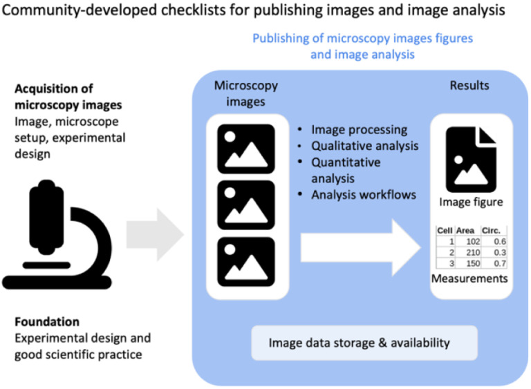
Images document scientific discoveries and are prevalent in modern biomedical research. Microscopy imaging in particular is currently undergoing rapid technological advancements. However for scientists wishing to publish the obtained images and image analyses results, there are to date no unified guidelines. Consequently, microscopy images and image data in publications may be unclear or difficult to interpret. Here we present community-developed checklists for preparing light microscopy images and image analysis for publications. These checklists offer authors, readers, and publishers key recommendations for image formatting and annotation, color selection, data availability, and for reporting image analysis workflows. The goal of our guidelines is to increase the clarity and reproducibility of image figures and thereby heighten the quality and explanatory power of microscopy data is in publications.


Figures

Figure 1.
Figure 1.
Scope of the checklists of image figure and image analysis requirements. The checklists present easy-to-use guidelines for publishing microscopy image figures and image analysis workflows.
Figure 2.
Figure 2.
Checklist for image publication including points to be addressed on image format, image colors and channels, image annotations, and image availability.
Figure 3.
Figure 3.
Image formatting may include (A) image cropping, rotation, and resizing, (B) image spacing in the figure, and (C) presenting several magnifications (zoom, inset) of images.
Figure 4.
Figure 4.
Image colors and channels. (A) Adjust brightness/contrast to achieve good visibility of the imaging signal. (B) Channel information should be annotated and visible to audiences (high contrast to background color, visible to color-blind audiences). (C) Image details are most unbiased in grayscale. (D) It is best practice to publish legends to color scales with images, and mandatory for pseudo-color scales.
Figure 5.
Figure 5.
Image Annotation. (A) Possible ways to provide scale information. (B) Features in images can be annotated with symbols, letters, or region-of- interest. (C) For advanced image publication, information on z-stack or voxel size, anatomical view, or camera settings such as pixel-dwell time, exposure time, or frame-time may be required.
Figure 6.
Figure 6.
Image Availability. (A) Currently, image data is often shared ‘upon request’. (B) More images along with the image metadata should be available for download in public databases, and in the future (C) also archived in dedicated, added-value databases, in which images are machine searchable or curated.
Figure 7.
Figure 7.
Image analysis. (A) An established workflow template is applied on new image data to produce a result (plot). (B) A new sequence of existing image analysis components is assembled into a novel workflow for a specific analysis (image segmentation). (C) Machine learning workflows learn specific tasks from data, and the resulting model is applied to obtain results.
Figure 8.
Figure 8.
Checklist for publication of image analysis workflows.

References

    1. Aaron J., Chew T.-L., 2021. A guide to accurate reporting in digital image processing - can anyone reproduce your quantitative analysis? J. Cell Sci. 134, jcs254151. 10.1242/jcs.254151 - DOI - PubMed
    1. Arganda-Carreras I., Kaynig V., Rueden C., Eliceiri K.W., Schindelin J., Cardona A., Sebastian Seung H., 2017. Trainable Weka Segmentation: a machine learning tool for microscopy pixel classification. Bioinforma. Oxf. Engl. 33, 2424–2426. 10.1093/bioinformatics/btx180 - DOI - PubMed
    1. Arzt M., Deschamps J., Schmied C., Pietzsch T., Schmidt D., Tomancak P., Haase R., Jug F., 2022. LABKIT: Labeling and Segmentation Toolkit for Big Image Data. Front. Comput. Sci. 4.
    1. Baker M., 2016. 1,500 scientists lift the lid on reproducibility. Nature 533, 452–454. 10.1038/533452a - DOI - PubMed
    1. Berginski M.E., Gomez S.M., 2013. The Focal Adhesion Analysis Server: a web tool for analyzing focal adhesion dynamics. F1000Research 2, 68. 10.12688/f1000research.2-68.v1 - DOI - PMC - PubMed

Publication types

LinkOut - more resources