[Preprint]. 2023 Feb 16:2023.02.16.528733.
doi: 10.1101/2023.02.16.528733.

Genetic manipulation of betta fish

Affiliations

Genetic manipulation of betta fish

Alec Palmiotti et al. bioRxiv. .

Update in

  • Genetic manipulation of betta fish.
    Palmiotti A, Lichak MR, Shih PY, Kwon YM, Bendesky A. Palmiotti A, et al. Front Genome Ed. 2023 Jul 21;5:1167093. doi: 10.3389/fgeed.2023.1167093. eCollection 2023. Front Genome Ed. 2023. PMID: 37545763 Free PMC article.

Abstract

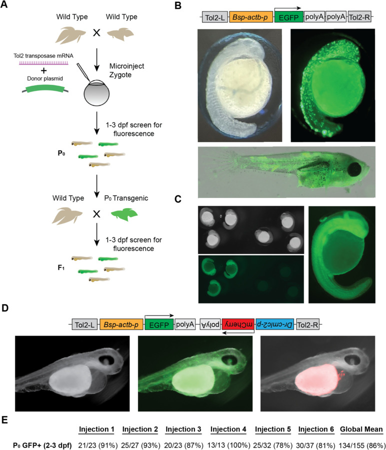
Betta splendens , also known as Siamese fighting fish or 'betta', are renowned for their astonishing morphological diversity and extreme aggressive behavior. Despite recent advances in our understanding of the genetics and neurobiology of betta, the lack of tools to manipulate their genome has hindered progress at functional and mechanistic levels. In this study, we outline the use of three genetic manipulation technologies, which we have optimized for use in betta: CRISPR/Cas9-mediated knockout, CRISPR/Cas9-mediated knockin, and Tol2-mediated transgenesis. We knocked out three genes: alkal2l, bco1l , and mitfa , and analyzed their effects on viability and pigmentation. Furthermore, we successfully knocked in a fluorescent protein into the mitfa locus, a proof-of-principle experiment of this powerful technology in betta. Finally, we used Tol2-mediated transgenesis to create fish with ubiquitous expression of GFP, and then developed a bicistronic plasmid with heart-specific expression of a red fluorescent protein to serve as a visible marker of successful transgenesis. Our work highlights the potential for the genetic manipulation of betta, providing valuable resources for the effective use of genetic tools in this animal model.


Conflict of interest statement

Conflict of Interest

The authors declare that the research was conducted in the absence of any commercial or financial relationships that could be construed as a potential conflict of interest

Figures

Figure 1.
Figure 1.. CRISPR/Cas9-mediated knockout in betta
(A) Experimental scheme for generating knockout betta. (B) Efficiency of knockout generation in P0, as determined by a T7EI assay. Numbers denote the fraction and percent of T7EI+ P0 individuals. (C) Schematic of alkal2l gene and the location of the gRNA target (top); wildtype DNA sequence in green and CRISPR/Cas9-induced mutations in magenta (middle); wildtype protein sequence (green shows the amino acids encoded by the wildtype sequence shown in the middle) and mutant sequence due to a frameshift (red shows the new mutant amino acids and * denotes a stop codon). (D) Boxplots showing proportion of blue (left) and red (right) pixels in alkal2l F3 crispants, according to genotype. P-values by Mann-Whitney test adjusted by Bonferroni correction. Boxes denote the interquartile range and whiskers the 5th and 95th percentiles, with a line at the median. (E) Representative images of betta individuals according to alkal2l genotype (left). Microscope images of the side of the body showing scales and the iridophore cover (center); the color of the microscope images appears different from the whole-body photographs on the left, due to the use of bright incident light under the microscope. Joint per-pixel quantification of hue, saturation and value by genotype (right).
Figure 2.
Figure 2.. CRISPR/Cas9-mediated knockin in betta
(A) Experimental scheme for generating knockin betta. (B) Brightfield (top), fluorescence only (middle) and merged images of wild type (non-injected) betta and P0 GFP+ betta at 24 hpf. (C) Efficiency of knockin generation across injection of three different clutches. Numbers denote the fraction and percent of GFP+ individuals; 95% of these GFP+ animals are also positive for a GFP-targeted locus junction PCR (see Results). (D) Schematic of mitfa gene and location of the gRNA target for insertion of GFP. (E) Brightfield (left), fluorescence only (center) and merge (right) of 24-hpf injected embryos with clear GFP expression (arrow) and no expression.
Figure 3.
Figure 3.. Tol2-mediated transgenesis in betta
(A) Experimental scheme for generating transgenic betta. (B) Schematic of pAB-1 actb:EGFP Tol2 plasmid (top), brightfield image of 24-hpf injected embryo (left) and fluorescent images of 24-hpf embryo (right) and ~30-dpf betta (bottom). (C) Brightfield (top left) and fluorescent bottom left, right) images of actb:EGFP F1 transgenics. (D) Schematic of pAB-16 Tol2 bicistronic plasmid (top) and images of 3-dpf injected embryos using bright field (left) and GFP filter (middle) and mCherry filter (right). (E) Efficiency of transgenic generation across injections of six different clutches. Numbers denote the fraction and percentage of GFP+ P0 individuals.

References

    1. Chen J., and Schedl T. (2021). A simple one-step PCR assay for SNP detection. MicroPublication Biol. 2021. doi: 10.17912/micropub.biology.000399. - DOI - PMC - PubMed
    1. Danner E., Bashir S., Yumlu S., Wurst W., Wefers B., and Kühn R. (2017). Control of gene editing by manipulation of DNA repair mechanisms. Mamm. Genome Off. J. Int. Mamm. Genome Soc. 28, 262–274. doi: 10.1007/s00335-017-9688-5. - DOI - PubMed
    1. Dehairs J., Talebi A., Cherifi Y., and Swinnen J. V. (2016). CRISP-ID: decoding CRISPR mediated indels by Sanger sequencing. Sci. Rep. 6, 28973. doi: 10.1038/srep28973. - DOI - PMC - PubMed
    1. Fan G., Chan J., Ma K., Yang B., Zhang H., Yang X., et al. (2018). Chromosome-level reference genome of the Siamese fighting fish Betta splendens, a model species for the study of aggression. GigaScience 7, giy087. doi: 10.1093/gigascience/giy087. - DOI - PMC - PubMed
    1. García-Nafría J., Watson J. F., and Greger I. H. (2016). IVA cloning: A single-tube universal cloning system exploiting bacterial In Vivo Assembly. Sci. Rep. 6, 27459. doi: 10.1038/srep27459. - DOI - PMC - PubMed

Publication types