[Preprint]. 2023 Feb 16:2023.02.15.528740.
doi: 10.1101/2023.02.15.528740.

E2F1 Mediates SOX17 Deficiency-Induced Pulmonary Hypertension

Affiliations

E2F1 Mediates SOX17 Deficiency-Induced Pulmonary Hypertension

Dan Yi et al. bioRxiv. .

Update in

  • E2F1 Mediates SOX17 Deficiency-Induced Pulmonary Hypertension.
    Yi D, Liu B, Ding H, Li S, Li R, Pan J, Ramirez K, Xia X, Kala M, Ye Q, Lee WH, Frye RE, Wang T, Zhao Y, Knox KS, Glembotski CC, Fallon MB, Dai Z. Yi D, et al. Hypertension. 2023 Nov;80(11):2357-2371. doi: 10.1161/HYPERTENSIONAHA.123.21241. Epub 2023 Sep 22. Hypertension. 2023. PMID: 37737027 Free PMC article.

Abstract

Rationale: Rare genetic variants and genetic variation at loci in an enhancer in SRY-Box Transcription Factor 17 (SOX17) are identified in patients with idiopathic pulmonary arterial hypertension (PAH) and PAH with congenital heart disease. However, the exact role of genetic variants or mutation in SOX17 in PAH pathogenesis has not been reported.

Objectives: To investigate the role of SOX17 deficiency in pulmonary hypertension (PH) development.

Methods: Human lung tissue and endothelial cells (ECs) from IPAH patients were used to determine the expression of SOX17. Tie2Cre-mediated and EC-specific deletion of Sox17 mice were assessed for PH development. Single-cell RNA sequencing analysis, human lung ECs, and smooth muscle cell culture were performed to determine the role and mechanisms of SOX17 deficiency. A pharmacological approach was used in Sox17 deficiency mice for therapeutic implication.

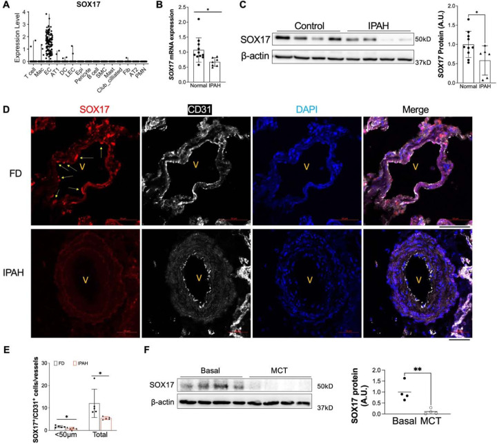
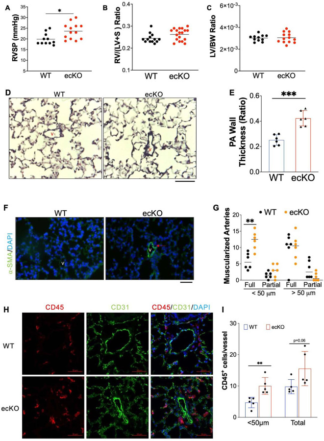
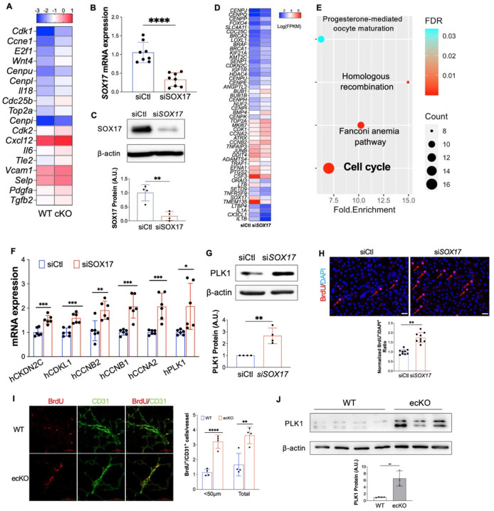
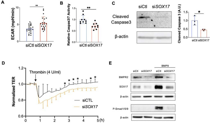
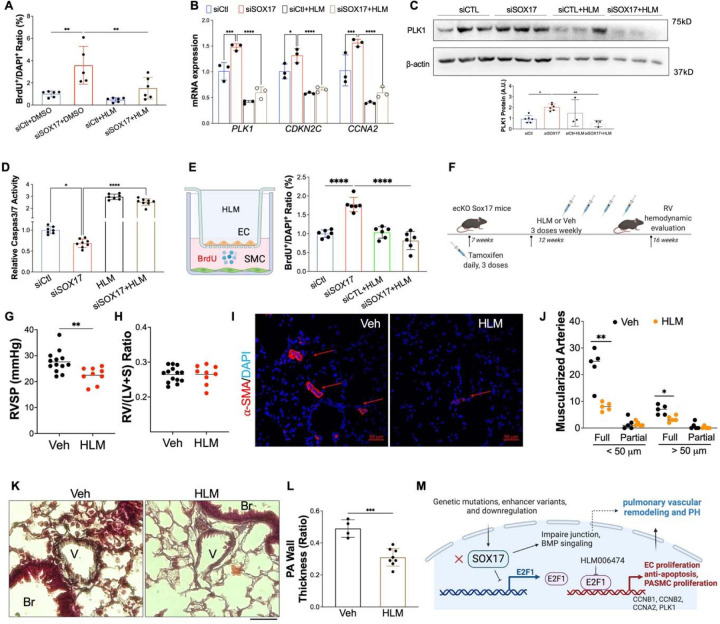
Measurement and main results: SOX17 expression was downregulated in the lungs and pulmonary ECs of IPAH patients. Mice with Tie2Cre mediated Sox17 knockdown and EC-specific Sox17 deletion developed spontaneously mild PH. Loss of endothelial Sox17 in EC exacerbated hypoxia-induced PH in mice. Loss of SOX17 in lung ECs induced endothelial dysfunctions including upregulation of cell cycle programming, proliferative and anti-apoptotic phenotypes, augmentation of paracrine effect on pulmonary arterial smooth muscle cells, impaired cellular junction, and BMP signaling. E2F Transcription Factor 1 (E2F1) signaling was shown to mediate the SOX17 deficiency-induced EC dysfunction and PH development.

Conclusions: Our study demonstrated that endothelial SOX17 deficiency induces PH through E2F1 and targeting E2F1 signaling represents a promising approach in PAH patients.


Figures

Figure 1.
Figure 1.. Downregulation of endothelial SOX17 in the patients with PAH.
(A) A violin plot showing SOX17 is restricted in the ECs of human lungs via scRNA-seq. Mac=macrophage; DC= dendritic cell; LEC=lymphatic EC; Epi=epithelium; SMC= smooth muscle cell; Fib=fibroblast; AT1 or AT2 = alveolar type 1 or 2 epithelium; PMN=neutrophils. (B) qRT-PCR analysis showed that SOX17 mRNA levels were downregulated in the sub-confluent PVECs isolated from IPAH patients. Each data point represents cells from one human subject including both male and female. (C) Western blotting demonstrated reduction of SOX17 protein expression in the IPAH PVECs. Each data point represents cells from one human subject including both male and female. (D, E) Immunostaining against SOX17 showing diminished SOX17 expression in the ECs of remodeling lesions from IPAH patients. Arrows indicate SOX17 positive ECs in non-PAH failed donors (FD). SOX17+/CD31+ cell number was quantified and normalized by vessels number. Each dot represents one subject. (F) SOX17 is decreased in the lungs of established PH rats at 4 weeks post MCT (33mg/kg subcutaneously) treatment. Student t test (B, C, E, F). *, P< 0.05; **, P< 0.01. A.U. = arbitrary units; Scale bar, 50 μm.
Figure 2.
Figure 2.. Tie2Cre mediated Sox17 deficiency induced PH and cardiac hypertrophy.
(A) Hemodynamic measurement showing that cKO Sox17 mice had increased right ventricular systolic pressure (RVSP) compared with Sox17f/f (WT) mice. (B and C) Cardiac dissection showed the upregulation of right heart and left heart hypertrophy in cKO mice compared with WT mice.. (D) Representative micrographs of Russell-Movat pentachrome staining showing increased medial thickness in Sox17 cKO mice compared with WT mice. (E) Quantification of pulmonary artery wall thickness. Wall thickness was calculated by the distance between internal wall and external wall divided by the distance between external wall and the center of lumen. (F and G) Muscularization of distal pulmonary vessels was markedly enhanced in Sox17 cKO mice compared with WT mice. Lung sections were immunostained with anti–α-SMA (green). Red arrow indicates a-SMA+ distal pulmonary vessels. α-SMA+ vessels were quantified in 20 field at 10X magnification per mouse (D) Student t test (A, B, C, E, G). *, P< 0.05; **, P< 0.01, ***, P< 0.001. Scale bar, 50 μm.
Figure 3.
Figure 3.. Endothelial SOX17 deficiency induced spontaneous mild PH.
(A) ecKO Sox17 mice exhibited increase of RVSP. (B and C) No change of RV and LV hypertrophy in ecKO Sox17 mice compared with WT mice. (D) Representative micrographs of Russell-Movat pentachrome staining showing increased medial thickness in ecKO Sox17 mice compared with WT mice. (E) Quantification of pulmonary artery wall thickness. Wall thickness was calculated by the distance between internal wall and external wall divided by the distance between external wall and the center of lumen. (F and G) Muscularization of distal pulmonary vessels was markedly enhanced in ecKO Sox17 mice compared with WT mice. Lung sections were immunostained with anti–α-SMA (green). Red arrow indicates a-SMA+ distal pulmonary vessels. α-SMA+ vessels were quantified in 20 field at 10X magnification per mouse. (H and I) Immunostaining against CD45 (Red) demonstrated that there was upregulated accumulation of inflammatory cells in the perivascular bed of ecKO Sox17 mice. Student t test (A, B, C, E, G, I). *, P< 0.05; **, P< 0.01, ***, P< 0.001. Scale bar, 50 μm.
Figure 4.
Figure 4.. Augmentation of PH by SOX17 deficiency in ECs under hypoxia.
(A) Hemodynamic measurement demonstrated that ecKO Sox17 mice exhibited increased of RVSP compared to WT mice under hypoxia condition. (B) RV dissection showing upregulation of RV hypertrophy in ecKO Sox17 mice compared to WT mice in response to hypoxia. (C and D) Quantification of Russell-Movat pentachrome staining showing thicker pulmonary artery walls and representative micrographs in ecKO Sox17 mice compared with WT mice in hypoxia condition. V=vessel, # indicates narrower vessel, * indicates occlusive vessel. Wall thickness was calculated by the distance between internal wall and external wall divided by the distance between external wall and the center of lumen. (E and F) Quantification of anti–α-SMA staining showing upregulation of muscularization of distal pulmonary artery wall and representative micrographs in ecKO Sox17 mice compared with WT mice in hypoxia condition. α-SMA+ vessels were quantified in 20 field at 10X magnification per mouse. Student t test (A, B, D and F). *, P< 0.05; **, P< 0.01, ***, P< 0.001. Scale bar, 50 μm.
Figure 5.
Figure 5.. Loss of SOX17 induced EC proliferation.
(A) scRNA transcriptomics showed that Sox17 deficiency ECs expressed higher levels of proliferation genes compared to WT ECs. scRNA-seq analysis was performed on the whole lung of WT and cKO mice. Lung ECs transcriptomics were analyzed. (B) qRT-PCR analysis showing efficient knockdown of SOX17 via siRNA against SOX17 in HPVECs. (C) siRNA against SOX17 markedly reduced SOX17 protein expression. (D) A representative heatmap of RNA-sequencing analysis of SOX17 knockdown in HPVECs. HPVECs were transfected with control siRNA (siCtl) or SOX17 siRNA for 48 hours. Equal amount of RNA from three replicates per group were pooled for RNA-seq. (E) KEGG pathway enrichment analysis of upregulated genes in SOX17 deficient lung ECs demonstrating that cell cycle pathway is the top upregulated signaling induced by loss of SOX17. (F) qRT-PCR analysis confirmed the upregulation of cell proliferation related genes including CKDN2C, CDKL1, CCNB2, CCNB1, CCNA2, and PLK1. (G) Western Blotting analysis demonstrated induction of PLK1 protein expression by SOX17 deficiency. (H) BrdU incorporation assay demonstrated increased of EC proliferation in SOX17 deficient HPVECs. At 48 hours post-transfection, HPVECs were starved in serum/growth factors free medium for 12 hours. BrdU was added in the medium at 4 hours prior to cells harvest. BrdU was stained with anti-BrdU antibodies. Red indicated BrdU positive cells. Nucleus were co-stained with DAPI. (I) In vivo BrdU incorporation assay showed upregulation of lung ECs proliferation in ecKO Sox17 mice during hypoxia condition. WT and ecKO Sox17 mice were incubated in hypoxia (10% O2) for 10 days. BrdU (25 mg/kg) was injected i.p. between day 7 to day 9. Lung sections were stained with anti-BrdU and anti-CD31. BrdU+/CD31+ cells were quantified. (J) Augmentation of cell proliferation marker PLK1 expression in the lung of ecKO Sox17 (ecKO) mice compared to WT mice. β-actin level was used as an internal control. Student t test (B, C, F, G, H, J). *, P< 0.05; **, P< 0.01. ***, P< 0.001. Scale bar, 50 μm.
Figure 6.
Figure 6.. SOX17 deficiency induced PASMC proliferation.
(A) A diagram showing the EC and SMCs co-culture model. (B and C) SOX17 deficiency in lung ECs promoted PASMCs proliferation assessed by Transwell co-culture and BrdU assay. PASMCs were seeded on the cover slides on the lower chamber. SOX17 deficiency or control HPVECs were seeded on the top chamber for 48 hours. PASMCs were starved overnight, then co-cultured with HPVECs. BrdU was added in the lower chamber at 8 hours prior to cells harvest. BrdU was stained with anti-BrdU antibodies. Red indicated BrdU positive cells. Nucleus were co-stained with DAPI. (D and E) In vivo BrdU incorporation assay showed upregulation of PASMCs proliferation in ecKO Sox17 mice during hypoxia condition. WT and ecKO Sox17 mice were incubated in hypoxia (10% O2) for 10 days. BrdU (25 mg/kg) was injected i.p. between day 7 to day 9. Lung sections were stained with anti-BrdU and anti- α-SMA. BrdU+/ α-SMA+ cells were quantified. (F) CellChat prediction using scRNA-seq dataset showed the upregulation of ligand and receptor pairs (Pdgfb-Pdgfra, Edn1-Ednra) in CKO mice. (G) ScRNA-seq analysis showed the increase of EC derived cytokines including Cxcl12, Edn1, Pdgfb, Pdgfd. Student t test (C and E). *, P< 0.05; **, P< 0.01. Scale bar, 50 μm.
Figure 7.
Figure 7.. Loss of endothelial SOX17 promoted EC dysfunction.
(A) Seahorse glycolytic assay showed that upregulation of Extracellular Acidification Rate (ECAR) levels in SOX17 deficient HPVECs compared to control cells. (B) SOX17 deficiency promoted anti-apoptotic phenotype of HPVECs during starvation assessed by Caspase 3/7 activities. (C) Western blotting analysis demonstrated reduction of cleaved Caspase 3 in SOX17 deficient HPVECs. (D) Impairment of endothelial barrier function in SOX17 deficient HPVECs. At 60 hours post-transfection, TER was monitored for up to 5 hours. Thrombin (4U/ml) was added to disrupt the cellular junction. (n=4). (E) Sox17 deficiency reduced BMPR2 expression and impaired BMPR2 activity via assessing P-Smad1/5/9 expression. Student t test (A-D). *, P< 0.05; **, P< 0.01.
Figure 8.
Figure 8.. E2F1 mediated SOX17 deficiency-induced dysfunction.
(A) iRegulon analysis demonstrated that FOXM1 and E2F1 are the top enriched transcriptional factors potentially governing cell cycle programming in SOX17 deficient HPVECs. (B) Upregulation of E2F1 protein expression by SOX17 knockdown. (C) Increased of E2F1 expression in the lung of ecKO Sox17 mice compared to WT mice. (D) qRT-PCR analysis showed that E2F1 siRNA markedly reduced E2F1 mRNA expression. (E) Western blotting analysis demonstrated that E2F1 protein was efficiently reduced by E2F1 siRNA compared to scramble siRNA. (F) QRT-PCR analysis demonstrated that E2F1 knockdown blocked the genes associated with proliferation including PLK1, CCNB1, and CCNB2 in the presence of SOX17 deficiency. (G) BrdU incorporation assay demonstrated that E2F1 knockdown normalized cell proliferation induced by loss of SOX17. (H) E2F1 knockdown restored EC apoptosis which was inhibited by SOX17 deficiency. Studies were repeated at least 3 times (B, D, F, G, H). Student t test (C, D and E). (I) A diagram shows that there are 3 putative SOX17 binding sites in the proximal promoter region of human E2F1 gene. (J) A representative map for pLV-E2F1P/Luc plasmid. (K) Loss of SOX17 increased E2F1 promoter activities assessed by luciferase assay. HPVECs were transfected with control of SOX17 siRNA for 12 hours, followed by infected with pLV-E2F1P/luc lentivirus for 48 hours. (L) A diagram showing that the SOX17 putative binding sites in E2F1 promoter/luciferase constructs were mutated. Purple highlight letters indicate mutated DNA sequences of the SOX17 putative binding sites in the E2F1 promoter. (M) Binding site 3 mutation blocked SOX17 deficiency-induced E2F1 promoter activation. MBS1/2/3 indicate mutated binding site 1/2/3. HPVECs were transfected with control of SOX17 siRNA for 12 hours, followed by infected with WT or mutated pLV-E2F1P/luc lentiviruses for 48 hours. Studies were repeated at least 3 times. One-way ANOVA with Tukey post hoc analysis (F, G and H). Student t test (K and M). *, P< 0.05; **, P< 0.01, ***, P< 0.001, ****, P< 0.0001.
Figure 9.
Figure 9.. Pharmacological inhibition of E2F1 reduced EC dysfunction and PH development in ecKO Sox17 mice.
(A) E2F1 inhibition reduced EC proliferation measured by BrdU incorporation assay. At 48 hours post-transfection of siRNA against SOX17 or control siRNA, HPVECs were treated with DMSO or HLM for 12 hours in serum/growth factors free medium. 2.5% FBS and BrdU were added in the medium at 4 hours prior to cells harvest. (B) qRT-PCR analysis demonstrated normalization of the expression of genes related to cell proliferation after E2F1 inhibition in HPVECs. At 48 hours post-transfection, HPVECs were treated with DMSO or HLM for 12 hours in serum/growth factors free medium. 2.5% FBS were added in the medium at 4 hours prior to RNA isolation. (C) E2F1 inhibition reduced cell proliferation marker PLK1 expression in SOX17 deficiency in HPVECs. (D) Pharmacological inhibition of E2F1 increased EC apoptosis in SOX17 deficient HPVECs. At 48 hours post-transfection, HPVECs were treated with DMSO or HLM for 12 hours in serum/growth factors free medium, followed by measurement of Caspase 3/7 activities. (E) A diagram showing the strategy of E2F1 inhibition in ecKO Sox17 mice. (F) RVSP was attenuated by E2F1 inhibition in ecKO Sox17 mice. (G) RV hypertrophy was not altered by E2F1 inhibition. (H and I) Muscularization of distal pulmonary arteries were reduced by E2F1 inhibition in ecKO Sox17 mice compared to vehicle. α-SMA+ vessels were quantified in 20 field at 10X magnification per mouse. (J and K) Pentachrome staining showed that E2F1 inhibition by HLM attenuated pulmonary wall thickness. Wall thickness was calculated by the distance between internal wall and external wall divided by the distance between external wall and the center of lumen. Studies were repeated at least 3 times (A, B, D). One-way ANOVA with Tukey post hoc analysis (A-D) and Student t test (F, G, I, K). *, P< 0.05; **, P< 0.01, ***, P< 0.001, ****, P< 0.0001. Scale bar, 50 μm.

References

    1. Corris PA, Seeger W. Call it by the correct name-pulmonary hypertension not pulmonary arterial hypertension: Growing recognition of the global health impact for a well-recognized condition and the role of the Pulmonary Vascular Research Institute. Am J Physiol - Lung Cell Mol Physiol 2020;318:L992–L994. - PubMed
    1. Moreira EM, Gall H, Leening MJG, Lahousse L, Loth DW, Krijthe BP, Kiefte-De Jong JC, Brusselle GG, Hofman A, Stricker BH, Ghofrani HA, Franco OH, Felix JF. Prevalence of pulmonary hypertension in the general population: The Rotterdam study. PLoS One 2015;10:1–12. - PMC - PubMed
    1. Austin ED, Newman JH, Loyd JE, Phillips JA. Heritable and Idiopathic Forms of Pulmonary Arterial Hypertension. Emery Rimoin’s Princ Pract Med Genet Genomics, Seventh Ed. Elsevier; 2020. p. 439–462.doi:10.1016/B978-0-12-812532-8.00016-1. - DOI
    1. Evans JDW, Girerd B, Montani D, Wang X-J, Galiè N, Austin ED, Elliott G, Asano K, Grünig E, Yan Y, Jing Z-C, Manes A, Palazzini M, Wheeler LA, Nakayama I, Satoh T, Eichstaedt C, Hinderhofer K, Wolf M, Rosenzweig EB, Chung WK, Soubrier F, Simonneau G, Sitbon O, Gräf S, Kaptoge S, Di Angelantonio E, Humbert M, Morrell NW. BMPR2 mutations and survival in pulmonary arterial hypertension: an individual participant data meta-analysis. Lancet Respir Med 2016;4:129–137. - PMC - PubMed
    1. Southgate L, Machado RD, Gräf S, Morrell NW. Molecular genetic framework underlying pulmonary arterial hypertension. Nat Rev Cardiol 2020;17:85–95. - PubMed

Publication types