[Preprint]. 2025 May 30:2023.02.14.528487.
doi: 10.1101/2023.02.14.528487.

CXCR6 promotes dermal CD8+ T cell survival and transition to long-term tissue residence

Affiliations

CXCR6 promotes dermal CD8+ T cell survival and transition to long-term tissue residence

Taylor A Heim et al. bioRxiv. .

Update in

Abstract

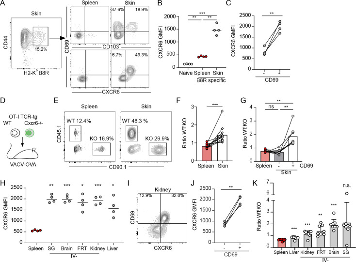
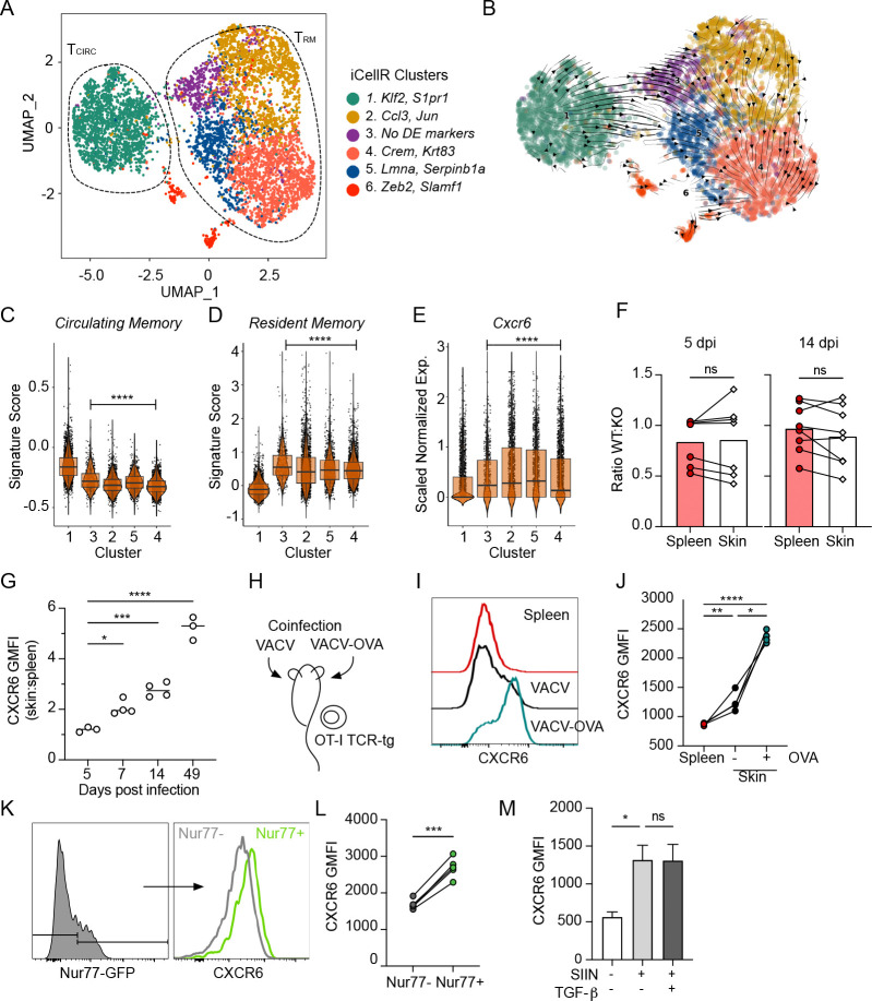
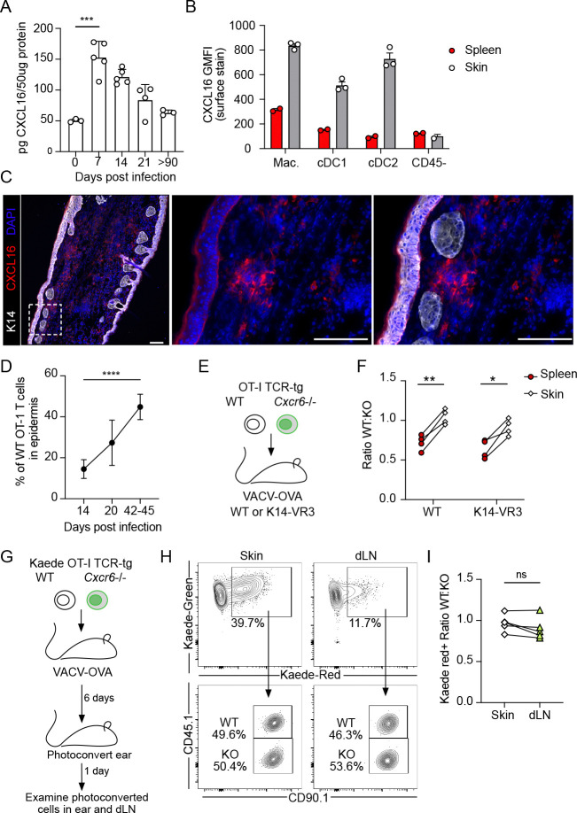
Tissue resident memory T cells (TRM) provide protection against local re-infection, and yet the interstitial signals necessary for their formation and persistence remain incompletely understood. Here we show that antigen-dependent induction of the chemokine receptor, CXCR6, is a conserved adaptation to peripheral tissue infiltration that promotes TRM formation after viral infection. Deficient TRM formation in the absence of CXCR6 was not explained by canonical trafficking as CXCR6 was not required for tissue entry, was dispensable for the early accumulation of antigen-specific CD8+ T cells in skin, and did not restrain their exit. Further, single cell sequencing indicated that Cxcr6 -/- CD8+ T cells were competent to acquire a transcriptional program of residence and TRM that formed were equally functional compared to their WT counterparts when reactivated greater than 100 days post primary infection. The reduced numbers observed at memory time points, where instead found to associate with impaired redox homeostasis and antioxidant capacity during the transition from effector to memory states. As such, Cxcr6 -/- CD8+ T cells exhibited increased rates of apoptosis in the dermis relative to controls, which led to reduced numbers of TRM in the epidermis at memory. CXCR6 therefore promotes the metabolic adaptation of T cells as they engage antigen in tissue to increase the probability of memory differentiation and long-term residence.


Conflict of interest statement

Competing interests: AWL reports consulting services for AGS Therapeutics. All other authors declare that they have no competing interests.

Figures

Figure 1.
Figure 1.. CXCR6 expression is a conserved feature of resident CD8+ T cells after viral infection.
(A) Representative flow of CD44+ vaccinia virus (VACV) specific (H2-KbB8R20–27) CD8+ T cells 28 days post infection (d.p.i.) (VACV, scarification) and expression of CD69, CD103, and CXCR6. (B, C) Quantification of CXCR6 geometric mean fluorescence intensity (GMFI) on CD44+CD8+ B8R-specific T cells from (A), as a function of location (B) and CD69 expression (C). (D) Experimental design where 15,000 wildtype (WT) OT-1 T cells (CD45.1+CD90.1) and 15,000 Cxcr6−/− OT-1 T cells (CD45.1CD90.1+) were co-transferred into naïve mice (CD45.1CD90.1) and the following day, ear skin was infected with VACV expressing OVA257–264 (VACV-OVA, scarification). (E) Representative flow plots at least 49 days post infection (d.p.i.). Gated on CD8+CD44+IV in the skin. (F) Quantification of the ratio of WT and Cxcr6−/− OT-1 T cells isolated from the spleen and ear skin from (E). (E and F) Data are cumulative from 3 experiments for a total of n=12. (G) Quantification of the ratio of WT and Cxcr6−/− 100–115 days post VACV infection stratified by CD69 expression. (H) CXCR6 expression on CD45.1+ OT-1 T cells in non-lymphoid tissues 31 d.p.i. with vesicular stomatitis virus expressing OVA (VSV-OVA) (gated on CD8+CD45.1+CD44+CD69+, and IV in all tissues except liver and spleen) (SG = salivary gland and FRT = Female reproductive tract). (I) Representative flow plot and (J) quantification of CXCR6 GMFI on IVCD45.1+ OT-1 T cells from kidney as a function of CD69 expression. (K) Quantification of the ratio of WT and Cxcr6−/− OT-1 T cells isolated 31 d.p.i. with VSV-OVA. Gated on CD8+CD44+IV in kidney, FRT, brain and salivary gland; CD8+CD44+ in liver and spleen. Data are cumulative of two experiments for a total of n=8. Bars represent average + SD. (A-G) Data are representative of at least two experiments with n=4 each. Statistical significance determined with one-way ANOVA (B, G, and H), repeated measures one-way ANOVA (K), and paired t test (C, F, and J). * p < 0.05, ** p < 0.01, *** p < 0.001, **** p < 0.0001
Figure 2.
Figure 2.. CXCR6 is required for optimal formation of TRM across non-lymphoid peripheral tissues.
(A) UMAP representation of single-cell RNA-seq data from extravascular (IV) OT-1 CD8+ T cells isolated out of skin 21 days post infection (d.p.i.) with vaccinia virus expressing OVA257–264 (VACV-OVA). Circulating (Tcirc) and resident (TRM) 5 memory T cells. Data represent a total of 7409 cells across all clusters. (B) Projected RNA velocity (scVelo) laid over UMAP. Individual iCellR clusters scored for a published (C) circulating memory, (D) resident memory, and (E) normalized Cxcr6 expression. (F) 15,000 wildtype (WT) and 15,000 Cxcr6−/− OT-1 T cells were co-transferred into naïve mice and the following day, skin was infected with VACV-OVA. Quantification of the ratio of WT and Cxcr6−/− OT-1 T cells in spleen and skin and 5 and 14 d.p.i. Data are cumulative from two experiments with n=4. (G) 10 Expression of CXCR6 by WT OT-1 T cells in skin relative to spleen over time. Data are representative of at least two experiments with n=3 or 4. (H) Experimental design where OT-1 T cells were transferred to naïve mice, which were subsequently infected on the left ear VACV-OVA and the right with VACV (scarification). (I) Representative histogram and quantification (J) of CXCR6 by OT-1 T cells in spleen or ear skin 14 d.p.i. Geometric mean fluorescence intensity (GMFI). Data are representative of at least two experiments with n=3 or 4. (K and L) Nurr77-GFP OT-1 T cells were transferred to naïve mice and the skin was infected with VACV-OVA the next day. (K) Representative histograms and (L) quantification of CXCR6 by Nurr77-GFP+ and Nurr77-GFP OT-1 T cells in skin 7 d.p.i. Data are representative of two experiments with n=5. (M) Expression of CXCR6 by effector CD8+ T cells from spleens of listeria monocytogenes (LM-OVA, 7 d.p.i) infected mice restimulated ex vivo with SIINFEKL and TGF-β. Bars represent average +SEM. Data are representative of two experiments with n=3. Cells in skin (F) are gated on IV-. Statistical significance determined using paired student’s t test (F and L) or one way ANOVA (G, J and M), or pairwise Wilcoxon rank test relative to cluster 1 (C-E). * p <0.05, ** p < 0.01, *** p < 0.001, **** p < 0.0001.
Figure 3.
Figure 3.. Dermal CXCL16 does not restrain T cell egress out of skin.
(A) Quantification of CXCL16 in vaccinia virus (VACV) infected skin at various days post infection (d.p.i) by scarification; n=3–5 per group. (B) Quantification of CXCL16 surface expression (GMFI) 12 d.p.i. on dermal and splenic myeloid cells including macrophages (Mac., CD11b+F480+), conventional dendritic cells type 1 (cDC1, CD11c+MHC-IIHIXCR1+) and type 2 (cDC2, CD11c+MHC-IIHISIRPA+). (C) Representative immunofluorescence images of skin 14 d.p.i. with VACV. Scale bar = 100μm. (C) The percentage of OT-1 T cells in the epidermis after infection with OVA257–264 -expressing VACV (VACV-OVA) calculated from immunofluorescent images. Data are cumulative of at least two experiments with n=5 (day 14) or n=6 (day 20 and 42–45). (E) Experimental design where 15,000 WT and 15,000 Cxcr6−/− OT-1 T cells were transferred to K14-VEGFR3-Ig mice and the following day infected with VACV-OVA via skin scarification. (F) Quantification of the ratio of WT and Cxcr6−/− OT-1 T cells in the spleen and skin of K14 VEGFR3-Ig mice 49 d.p.i. with VACV-OVA. Data are representative from at least two experiments with n=4. (G) Experimental design where congenically distinct 15,000 WT Kaede+ OT-1 T cells and 15,000 Cxcr6−/− Kaede+ OT-1 T cells were co-transferred to C57Bl/6J mice and infected the following day with VACV-OVA via skin scarification. Kaede expressing cells in skin were photoconverted 6 d.p.i. and harvested 1 day later from skin and draining lymph node (dLN). (H) Representative flow of skin and dLN. (I) The ratio of photoconverted WT Kaede red+ to photoconverted Cxcr6−/− Kaede red+ in skin and dLN following photoconversion. Data are representative from at least two experiments with n=5. Statistical significance determined using one-way ANOVA (A,D) or paired student’s t test (F,I). Error bars represent SD. * p < 0.05, ** p < 0.01, *** p < 0.001, **** p < 0.0001
Figure 4.
Figure 4.. Cxcr6−/− T cells fail to fully acquire a residence transcriptional program and display signs of metabolic dysfunction and apoptosis.
(A) UMAP showing merged wildtype (WT) and Cxcr6−/− OT-1 T cells isolated from skin 21 days post infection (d.p.i.) with vaccinia virus expressing OVA257–264 (VACV-OVA). Resident (TRM) and circulating (TCIRC) clusters defined as in Fig 2. (B) Cell density plots for WT and Cxcr6−/− OT-I T cells overlaid on the merged UMAP. (C) Cells in TCIRC cluster and TRM clusters scored for published circulating memory (left) or resident memory (right) gene signatures. (D) Volcano plot of differentially expressed genes (DEG) between WT and Cxcr6−/− cells in combined TRM clusters (–5). (E) Pathway analysis of DEG between WT and Cxcr6−/− cells in TRM clusters. p value calculated by hypergeometric distribution test. (F) Human Base analysis of gene modules downregulated in Cxcr6−/− TRM relative to WT. (G) Mito Deep Red GMFI and (H) MitoSox Red GMFI in WT and Cxcr6−/− OT-I T cells 14 d.p.i. (I) Ex vivo uptake of Zombie red viability dye 21 d.p.i. Statistical significance determined using paired student’s t test (G-I) or pairwise Wilcoxon rank test (C). * p < 0.05, *** p < 0.001, **** p < 0.0001.
Figure 5.
Figure 5.. CXCR6 promotes dermal T cell survival during memory formation in infected skin.
(A) Representative flow plots and (B) quantification of ex vivo uptake of ZombieRed viability dye in wildtype (WT) and Cxcr6−/− (KO) OT-1 T cells in the dermis and epidermis 14 days post infection (d.p.i) with vaccinia virus expressing OVA257–264 (VACV-OVA). Data are representative of at least two experiments with n=4. (C) Representative immunofluorescence image and (D) quantification of WT and Cxcr6−/− (KO) OT-1 T cells in skin at 14 d.p.i. with VACV-OVA. (E) Representative immunofluorescence and (F) quantification of WT and Cxcr6−/− (KO) OT-1 T cells in skin 42–45 d.p.i. with VACV-OVA. White line delineates the epidermis. Scale bar = 100μm. Data are cumulative of two experiments with n=5 (C,D) or n=6 (E,F). (G) Representative flow plots gated on live CD44+IV WT and Cxcr6−/− OT-I T cells in VACV-OVA immune mice restimulated with cognate peptide (SIINFEKL; 5 hrs). (H) Percent and (I) number of IFNg producing cells from G. Statistical significance determined using paired student’s t test. * p < 0.05, **p<0.01, *** p < 0.001.

References

    1. Bromley S. K., Yan S., Tomura M., Kanagawa O., and Luster A. D.. 2013. Recirculating Memory T Cells Are a Unique Subset of CD4+ T Cells with a Distinct Phenotype and Migratory Pattern. J. Immunol. 190: 970–976. - PMC - PubMed
    1. Mackay L. K., Rahimpour A., Ma J. Z., Collins N., Stock A. T., Hafon M.-L., Vega-Ramos J., Lauzurica P., Mueller S. N., Stefanovic T., Tscharke D. C., Heath W. R., Inouye M., Carbone F. R., and Gebhardt T.. 2013. The developmental pathway for CD103+CD8+ tissue-resident memory T cells of skin. Nat. Immunol. 14: 1294–1301. - PubMed
    1. Skon C. N., Lee J.-Y., Anderson K. G., Masopust D., Hogquist K. A., and Jameson S. C.. 2013. Transcriptional downregulation of S1pr1 is required for establishment of resident memory CD8+ T cells. Nat. Immunol. 14: 1285–1293. - PMC - PubMed
    1. Mackay L. K., Braun A., Macleod B. L., Collins N., Tebartz C., Bedoui S., Carbone F. R., and Gebhardt T.. 2015. Cutting edge: CD69 interference with sphingosine-1-phosphate receptor function regulates peripheral T cell retention. J. Immunol. Baltim. Md 1950 194: 2059–2063. - PubMed
    1. Kumar B. V., Ma W., Miron M., Granot T., Guyer R. S., Carpenter D. J., Senda T., Sun X., Ho S.-H., Lerner H., Friedman A. L., Shen Y., and Farber D. L.. 2017. Human Tissue-Resident Memory T Cells Are Defined by Core Transcriptional and Functional Signatures in Lymphoid and Mucosal Sites. Cell Rep. 20: 2921–2934. - PMC - PubMed

Publication types