[Preprint]. 2023 Feb 15:2023.02.15.528675.
doi: 10.1101/2023.02.15.528675.

Temporal Notch signaling regulates mucociliary cell fates through Hes-mediated competitive de-repression

Affiliations

Temporal Notch signaling regulates mucociliary cell fates through Hes-mediated competitive de-repression

Magdalena Maria Brislinger-Engelhardt et al. bioRxiv. .

Abstract

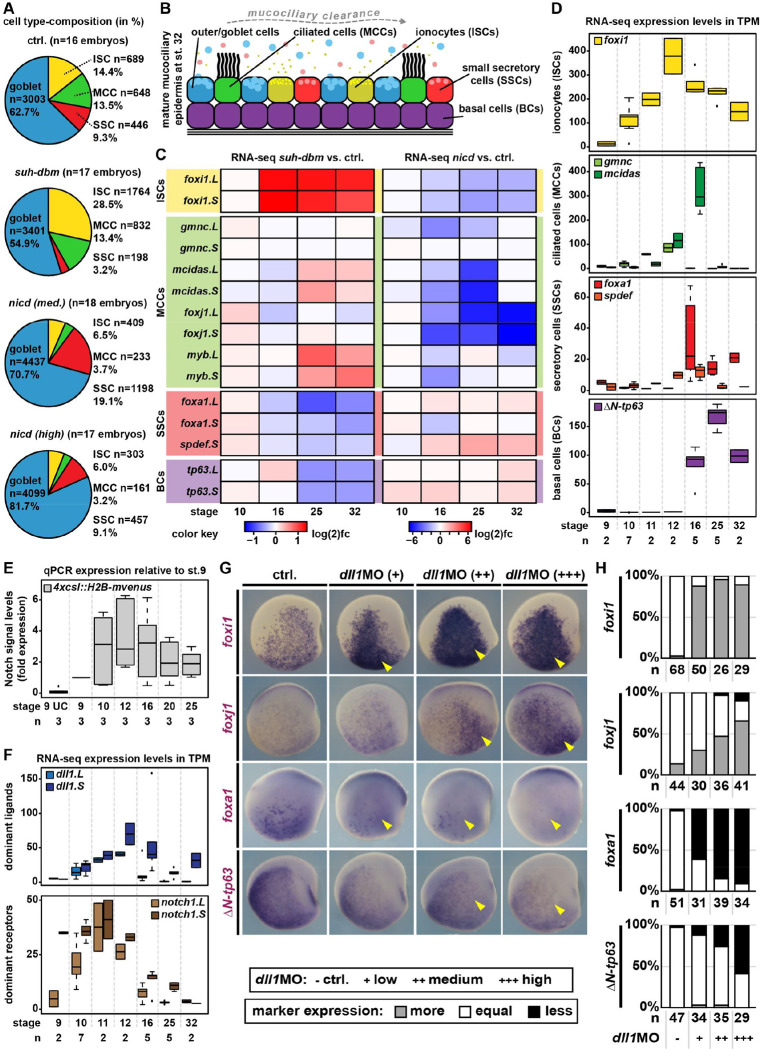
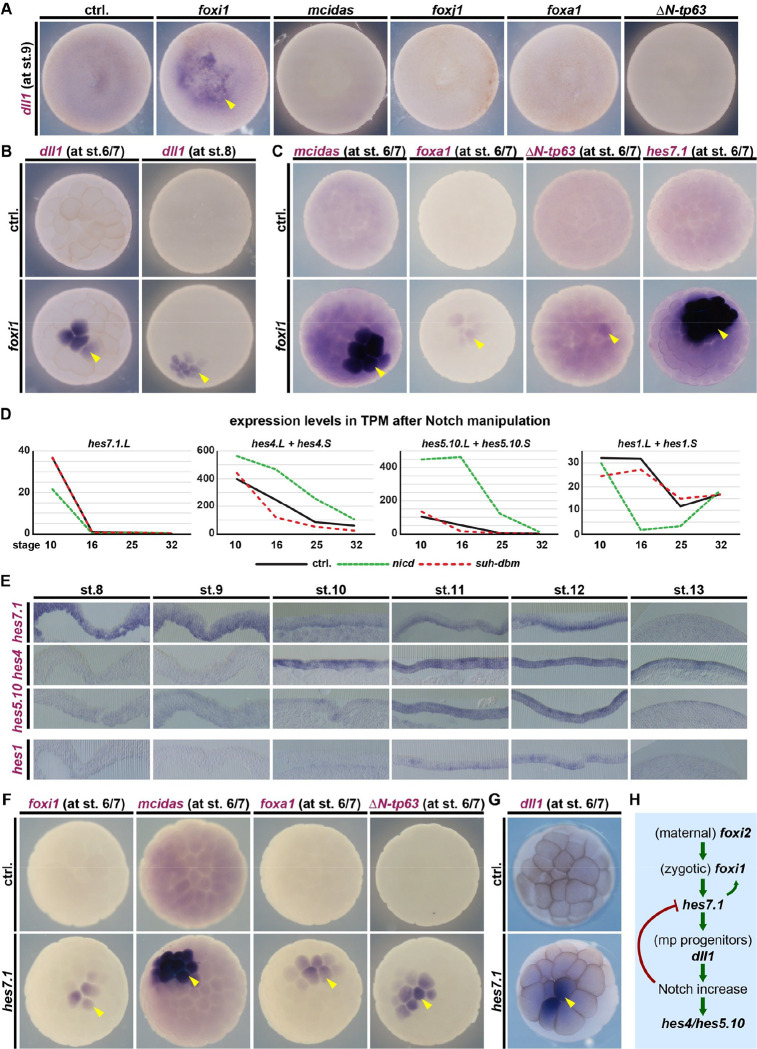
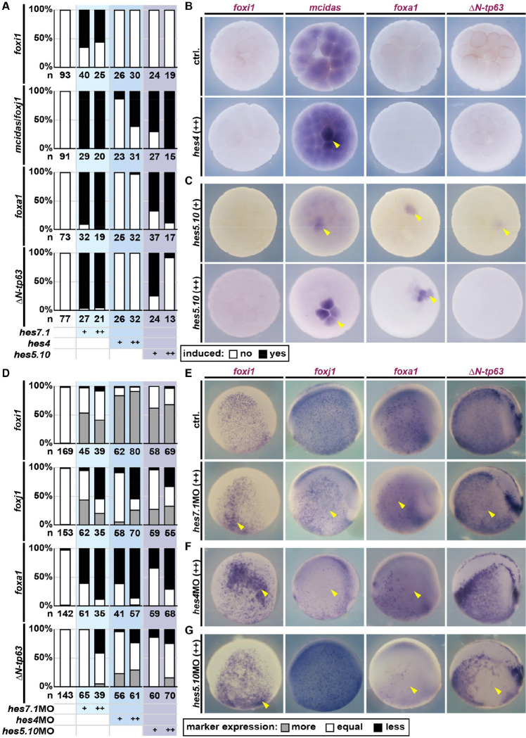
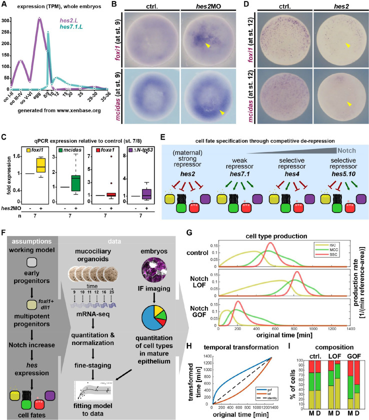
Tissue functions are determined by the types and ratios of cells present, but little is known about self-organizing principles establishing correct cell type compositions. Mucociliary airway clearance relies on the correct balance between secretory and ciliated cells, which is regulated by Notch signaling across mucociliary systems. Using the airway-like Xenopus epidermis, we investigate how cell fates depend on signaling, how signaling levels are controlled, and how Hes transcription factors regulate cell fates. We show that four mucociliary cell types each require different Notch levels and that their specification is initiated sequentially by a temporal Notch gradient. We describe a novel role for Foxi1 in the generation of Delta-expressing multipotent progenitors through Hes7.1. Hes7.1 is a weak repressor of mucociliary genes and overcomes maternal repression by the strong repressor Hes2 to initiate mucociliary development. Increasing Notch signaling then inhibits Hes7.1 and activates first Hes4, then Hes5.10, which selectively repress cell fates. We have uncovered a self-organizing mechanism of mucociliary cell type composition by competitive de-repression of cell fates by a set of differentially acting repressors. Furthermore, we present an in silico model of this process with predictive abilities.

Keywords: Xenopus; airway epithelium; basal cells; development; mathematical modeling; mucus; multi-ciliated cells.


Figures

Figure 1:
Figure 1:. Cell fate specification in response to a temporal Notch gradient
A: Quantification of epidermal cell types after Notch manipulations and immunofluorescent staining at st. 32. B: Schematic representation of mucociliary epidermal cell types. C: Changes in cell type marker expression relative to stage-matched controls after manipulation of Notch signaling in Xenopus mucociliary organoids (RNA-seq). D,F: Expression levels of indicated genes in unmanipulated mucociliary organoids across different stages of development (RNA-seq). E: Notch reporter activity in mucociliary organoids relative to 4xcsl::H2B-mvenus injected st. 9 samples over time (qPCR). G: Lateral view, WMISH for indicated transcripts at st. 16/17 in controls and after knockdown of dll1. +=low dose, ++=medium dose, +++=high dose. H: Quantification of results depicted in G. Expression levels were scored more, equal, less expression on the injected vs. uninjected side.
Figure 2:
Figure 2:. Foxi1 induces multipotent ligand-expressing progenitors via hes7.1
A-C, F,G: Animal view, WMISH. A: dll1 at st. 9 in controls and after overexpression of master cell type inducers. Only foxi1 induces dll1 expression (yellow arrowhead). B: dll1 at st. 6/7 and 8 in controls and after foxi1 (induction=yellow arrowhead). C: Marker gene expression at st. 6/7 in controls and after foxi1 (induction=yellow arrowhead). D: Expression levels of indicated hes transcripts in unmanipulated mucociliary organoids (black line) and after Notch manipulations (loss=red line; gain=green line) across different stages of development (RNA-seq). E: Epidermis sections, WMISH for indicated hes transcripts from st. 8 to st. 13. F,G: Marker gene expression at st. 6/7 in controls and after hes7.1 (induction=yellow arrowhead). H: Schematic summary of Notch, hes and dll1 regulation in multipotent progenitors.
Figure 3:
Figure 3:. Hes factors regulate mucociliary cell fates
A: Quantification of results depicted in B,C. B,C: Animal view, WMISH. Marker gene expression at st. 6/7 in controls and after overexpression of indicated hes, +=low dose; ++=high dose (induction=yellow arrowhead). D: Quantification of results depicted in E-G. Expression levels were scored more, equal, less expression on the injected vs. uninjected side. E-G: Lateral view, WMISH. Marker gene expression at st. 16/17 in controls and after knockdown of indicated hes, +=low dose; ++=high dose (changes in expression=yellow arrowhead).
Figure 4:
Figure 4:. Competitive de-repression and modeling of mucociliary cell fate decisions
A: Expression levels of hes2.L and hes7.1.L in whole Xenopus eggs and embryos during early development. (RNA-seq.) B: Animal view, WMISH. Marker gene expression at st. 9 in controls and after knockdown of hes2. (induction=yellow arrowhead). C: Induction of mucociliary marker genes after knockdown of hes2 at st. 7/8 in whole embryos. (qPCR). D: Ventral view, WMISH. Marker gene expression at st. 12 in controls and after overexpression of hes2. (loss of expression=yellow arrowhead). E: Schematic summary of Hes-mediated competitive de-repression of cell fates in Xenopus. F: Schematic representation of minimal-component model building. G-I: In silico modeling of the cell fate specification process. G: Cell type production rates over time (control), and the effect of deceleration (LOF) or acceleration (GOF) on the production rates of ISCs, MCCs, and SSCs. H: The in-silico model could explain Notch loss- (LOF) and gain-of-function (GOF) experiments by deceleration (LOF) or acceleration (GOF) of the whole cell specification process via the depicted nonlinear transformation of the time axis. I: Comparison of modeled [M] and experimentally determined [D] cell type ration of ISCs (yellow), MCCs (green) and SSCs (red).

Similar articles

References

    1. Briggs J. A., Weinreb C., Wagner D. E., Megason S., Peshkin L., Kirschner M. W. and Klein A. M. (2018). The dynamics of gene expression in vertebrate embryogenesis at single-cell resolution. Science (80-. ). 360, eaar5780. - PMC - PubMed
    1. Cha S. W., McAdams M., Kormish J., Wylie C. and Kofron M. (2012). Foxi2 is an animally localized maternal mRNA in xenopus, and an activator of the zygotic ectoderm activator foxi1e. PLoS One 7, 1–10. - PMC - PubMed
    1. Community T. G. (2022). The Galaxy platform for accessible , reproducible and collaborative biomedical analyses : 2022 update. Nucleic Acids Res. 50, 345–351. - PMC - PubMed
    1. Deblandre G. A., Wettstein D. A., Koyano-Nakagawa N. and Kintner C. (1999). A two-step mechanism generates the spacing pattern of the ciliated cells in the skin of Xenopus embryos. Development 126, 4715–4728. - PubMed
    1. Dobin A., Davis C. A., Schlesinger F., Drenkow J., Zaleski C., Jha S., Batut P., Chaisson M. and Gingeras T. R. (2013). STAR: ultrafast universal RNA-seq aligner. Bioinformatics 29, 15–21. - PMC - PubMed

Publication types