Review
. 2023 Apr;45(4):e2300004.
doi: 10.1002/bies.202300004. Epub 2023 Feb 24.

Vasa, a regulator of localized mRNA translation on the spindle

Affiliations
Review

Vasa, a regulator of localized mRNA translation on the spindle

Paola Alejandra Sundaram Buitrago et al. Bioessays. 2023 Apr.

Abstract

Localized mRNA translation is a biological process that allows mRNA to be translated on-site, which is proposed to provide fine control in protein regulation, both spatially and temporally within a cell. We recently reported that Vasa, an RNA-helicase, is a promising factor that appears to regulate this process on the spindle during the embryonic development of the sea urchin, yet the detailed roles and functional mechanisms of Vasa in this process are still largely unknown. In this review article, to elucidate these remaining questions, we first summarize the prior knowledge and our recent findings in the area of Vasa research and further discuss how Vasa may function in localized mRNA translation, contributing to efficient protein regulation during rapid embryogenesis and cancer cell regulation.

Keywords: DDX4; Vasa; asymmetric cell division; embryonic cells; localized translation; sea urchin; spindle.


Conflict of interest statement

CONFLICT OF INTEREST

The authors declare no conflict of interest.

Figures

Fig. 1.
Fig. 1.. Increased mRNA translation mediated by Vasa on the micromere-side of the spindle contributes to micromeres’ fates and their organizers’ functions through an asymmetric cell division, while the ectopic localization of Vasa facilitates ectopic protein synthesis in the cell, resulting in the developmental failure of the embryos.
Animal blastomeres undergo a symmetric cell division, while vegetal blastomeres undergo an asymmetric cell division at the 16-cell stage of the sea urchin embryo. A, Vasa (purple) becomes enriched equally on both sides of the spindles both in the animal and vegetal blastomeres at metaphase. B-C, Vasa in the animal blastomeres continues to be distributed evenly over the spindle, while that in the vegetal blastomeres becomes asymmetric toward the micromere-side of the spindle from anaphase to telophase, resulting in the Vasa accumulation in the micromeres. D, Vasa remains enriched in micromeres independent of the cell cycle, while it disperses into the cytoplasm during the S-phase in other blastomeres. Vasa enrichment comes back at perinuclear at the next M-phase entry. E, Vasa remains enriched in the germline, while largely cleared from the somatic lineages by the gastrula stage. F-H, A hypothetical model for Vasa’s function on the spindle during asymmetric cell division. Vasa granules help assemble ribosomes and mRNAs for active translation (F). Vasa protein increases on the micromere side of the spindle through the translation of its own mRNA, which facilitates the general protein synthesis more on the micromere side of the spindle (G). This results in the enriched and distinct molecular status of micromeres immediately upon the cytokinesis completion, contributing to the rapid lineage segmentation of micromeres (H). I, Forcing the Vasa localization at the ectopic place (e.g. the membrane) induces ectopic protein synthesis, which results in developmental failure such as no gastrulation. This suggests the proper localization of Vasa protein on the spindle is critical for embryonic development.
Fig. 2.
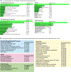
Fig. 2.. Enrichment pathway analyses of Vasa RNA-IP-seq datasets in Drosophila germ cells (A) and DDX4-overexpression proteomics datasets in human SCLC cells (B).
A, A list of genes was obtained from Liu et al., 2009 [38] and processed for the analyses using the FlyEnrichr to obtain the diagrams shown. Different color codes are given based on the adjusted P-value listed on the right to help visualization. B, A list of proteins upregulated in the DDX4-overexpressing group compared to the control (LUC) group was obtained from Noyes et al., 2023 [37] and processed for the analyses using the GSEA(https://www.gsea-msigdb.org/gsea/index.jsp) to obtain the tables shown.
Fig. 3.
Fig. 3.. Dynamic change of Vasa granule formation and localization during the cell cycle of the sea urchin embryo.
Vasa (magenta) accumulates as small granules at perinuclear prior to prophase, enters into the nucleus area upon the nuclear envelope breakdown, and forms visibly larger condensates around the chromosomes (blue) and the spindle (green) during the M-phase. These granules fade away toward the telophase and appear to disperse into the cytoplasm upon the M-phase exit, and come back at perinuclear for the next cell cycle.
Fig. 4.
Fig. 4.. Predicted functional domains of Vasa protein (left) and the sequence comparison of the conserved C-terminal region across organisms (right).
Diagrams are modified from Noyes et al., 2023.[37]
Fig. 5.
Fig. 5.. Dynamics of Vasa granules on the spindle.
A, The wildtype Vasa forms visible granules over the spindle (left, arrows), while Vasa-C3 mutant in which the last three amino acids were mutated failed to form granules or localize on the spindle (right, arrows). Green, Vasa-GFP; Magenta, 2xmCherry-EMTB (microtubule marker). B, An example of the solid status of Vasa granules on the spindle. Kaede-Vasa fluoresces in green in the intact form, while it changes color to red fluorescence upon photoconversion with UV light. Kaede-Vasa was photoconverted at the macromere side of the spindle (bracket “Macro-side”) during asymmetric cell division. The photoconverted Kaede-Vasa granules (arrows) did not move toward the micromere side yet remained at the original location through the M-phase, suggesting no protein translocation within the spindle. Green, unphotoconverted Kaede-Vasa; Magenta, photoconverted Kaede-Vasa. These images are modified from Fernandez-Nicholas et al., 2022.[12] Scale bars = 20 μm.

Similar articles

Cited by

References

    1. Gasparski AN, Mason DE, Moissoglu K, & Mili S (2022). Regulation and outcomes of localized RNA translation. Wiley Interdiscip Rev RNA, 13(6), e1721. 10.1002/wrna.1721 - DOI - PMC - PubMed
    1. Das S, Vera M, Gandin V, Singer RH, & Tutucci E (2021). Intracellular mRNA transport and localized translation. Nat Rev Mol Cell Biol, 22(7), 483–504. 10.1038/s41580-021-00356-8 - DOI - PMC - PubMed
    1. Waldron A, & Yajima M (2020). Localized translation on the mitotic apparatus: A history and perspective. Dev Biol, 468(1–2), 55–58. 10.1016/j.ydbio.2020.09.010 - DOI - PMC - PubMed
    1. Gross PR, Philpott DE, & Nass S (1958). The fine-structure of the mitotic spindle in sea urchin eggs. J Ultrastruct Res, 2(1), 55–72. 10.1016/s0022-5320(58)90047-9 - DOI - PubMed
    1. Gross PR, & Cousineau GH (1963). Synthesis of Spindle-Associated Proteins in Early Cleavage. J Cell Biol, 19, 260–265. 10.1083/jcb.19.1.260 - DOI - PMC - PubMed

Publication types

MeSH terms