. 2023 Aug;19(8):3537-3554.
doi: 10.1002/alz.12970. Epub 2023 Feb 24.

Pathogenesis of Alzheimer's disease: Involvement of the choroid plexus

Affiliations

Pathogenesis of Alzheimer's disease: Involvement of the choroid plexus

Maria Čarna et al. Alzheimers Dement. 2023 Aug.

Abstract

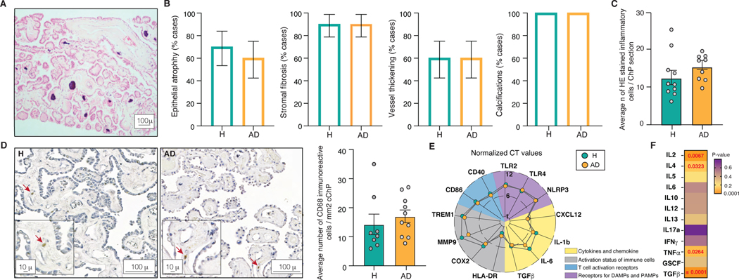
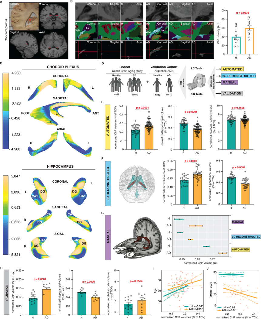
The choroid plexus (ChP) produces and is bathed in the cerebrospinal fluid (CSF), which in aging and Alzheimer's disease (AD) shows extensive proteomic alterations including evidence of inflammation. Considering inflammation hampers functions of the involved tissues, the CSF abnormalities reported in these conditions are suggestive of ChP injury. Indeed, several studies document ChP damage in aging and AD, which nevertheless remains to be systematically characterized. We here report that the changes elicited in the CSF by AD are consistent with a perturbed aging process and accompanied by aberrant accumulation of inflammatory signals and metabolically active proteins in the ChP. Magnetic resonance imaging (MRI) imaging shows that these molecular aberrancies correspond to significant remodeling of ChP in AD, which correlates with aging and cognitive decline. Collectively, our preliminary post-mortem and in vivo findings reveal a repertoire of ChP pathologies indicative of its dysfunction and involvement in the pathogenesis of AD. HIGHLIGHTS: Cerebrospinal fluid changes associated with aging are perturbed in Alzheimer's disease Paradoxically, in Alzheimer's disease, the choroid plexus exhibits increased cytokine levels without evidence of inflammatory activation or infiltrates In Alzheimer's disease, increased choroid plexus volumes correlate with age and cognitive performance.

Keywords: Alzheimer's disease; aging; cerebrospinal fluid; choroid plexus; pathology.


Conflict of interest statement

Conflicts of interest

No competing interests.

Figures

Figure 1.
Figure 1.. CSF in aging and AD
(A) Characteristics of the CSF protein database from the Emory Goizueta ADRC cohort. (B) The most significant Ingenuity pathways based on the CSF proteins changed by aging (p≤0.05). (C) The most significant Ingenuity pathways based on the CSF proteins changed by aging in healthy individuals versus AD (p≤0.05). (D) Volcano plot showing significantly changed CSF proteins in AD compared with healthy individuals in 45–55 (n=29), 56–65 (n=97), 66–75 (n=133) and 76–90 (n=38) year-old age groups (p≤0.05, NS: not significant). (E) The most significant Ingenuity pathways based on the CSF proteins changed by aging in AD compared with healthy individuals in different age groups (p≤0.05). (F) Sankey diagram showing significantly changed inflammatory pathways in AD compared with healthy individuals across different age groups (p≤0.05).
Figure 2.
Figure 2.. CSF in AD and other conditions
(A) Characteristics of the Czech Brain Aging Study cohort. (B) Heatmap representing hierarchically clustered CSF proteins identified by two independent mass spectrometers and protocols (A and B) in MCI, ALymeP and ALS compared with AD (p≤ 0.05 FDR adjusted t-test). (C) The most common significantly changed Reactome pathways in MCI, ALymeP and ALS compared with AD (p≤0.05). (D) Circular netplots showing significantly changed inflammatory CSF proteins and pathways in AD compared with ALymeP and ALS (p≤0.05).
Figure 3.
Figure 3.. Structure and the inflammatory status of the ChP in AD
(A) Representative H&E stained FFPE section of the ChP. (B) Percentage of healthy individuals and AD patients showing epithelial atrophy, stromal fibrosis, vessel thickening and calcifications in the ChP. 1 technical replicate ± s.e.m.; two-sample t-test (p≤0.05). (C) Average number of inflammatory cells in H&E stained ChP from healthy individuals and AD patients. Mean number of cells per 6 high-power microscopy fields per section ± s.e.m.; two-sample t-test (p≤0.05). (D) Anti-CD68 antibody labelled cells in the ChP of healthy individuals and AD patients. Mean number of cells per 10 high-power microscopy fields per section ± s.e.m.; two-sample t-test (p≤0.05). (E) Expression levels of markers of the activation of the ChP immune cells in healthy individuals and AD. The lines crossing central, middle and outermost circle represent distances corresponding to normalized CT values of 1, 6 and 12, respectively. (F) ELISArray measured ChP cytokine levels in healthy individuals and AD. Mean of 3 technical replicates per sample ± s.e.m.; two-sample t-test (p≤0.05).
Figure 4.
Figure 4.. Aberrant accumulation of CSF resident proteins in the ChP in AD
(A) Levels of CSF resident proteins in the ChP of healthy individuals and AD patients. Skyline iRT retention time prediction algorithm showing identical chromatogram retention times between proteotypic peptides used to quantify ChP proteins (right) and stable isotopically labelled synthetic peptides (left). Mean of 6 technical replicates per sample ± s.e.m.; two-sample t-test (p≤0.05). (B) Mean intensities of NPC2 and TTR in the scanned ChP of heathy individuals and AD patients (magnification bar 100 μ). Mean of 90 technical replicates per sample ± s.e.m.; two-sample t-test (p≤0.05). (C) Mean NPC2 and TTR intensities in the confocal images of the ChP epithelium and stroma in healthy individuals and AD (magnification bar 10 μ). Mean of 3–12 technical replicates ± s.e.m.; repeated measures (p≤0.05). (D) Levels of ChP gangliosides in healthy individuals and AD. Mean of 6 technical replicates per sample ± s.e.m.; two-sample t-test (p≤0.05).
Figure 5.
Figure 5.. Increased intensity, remodelling and volume of ChP in AD
(A) A coronal view of post-mortem ChP in the right lateral ventricles (red asterisk) and T1-weighted sequence derived positions of ChP in coronal, sagittal and axial views (in red). (B) Coronal, sagittal and axial views of ChP representative of healthy individuals and AD patients visualized using T1-weighted sequence (top), FreeSurfer brain structure map (middle) and following ChP segmentation (bottom). Normalized ChP intensities in healthy individuals and AD. Individual measurements ± s.e.m.; two-sample t-test (p≤0.05). (C) Mean changes in the shape of the 3D representations of the ChP and hippocampi in AD compared with healthy individuals. Single measurements ± s.e.m.; two-sample t-test (p≤0.05). (D) Experimental design of the ChP volumetry study. (E) Automated measurements of normalized mean ChP, hippocampal and cerebellar cortical volumes in healthy individuals and AD. Individual measurements ± s.e.m.; two-sample t-test (p≤0.05). (F) 3D reconstruction of the ChP (red) within the ventricular system (blue) in the brain. Normalized mean 3D representation-derived ChP and hippocampal volumes in healthy individuals and AD. Single measurements ± s.e.m.; two-sample t-test (p≤0.05). (G) 3D representation of manually traced ChP (in red). 95% confidence intervals of normalized automated, 3D reconstructed and manually traced ChP volumes in healthy individuals and AD. (H) Normalized mean ChP, hippocampal and cerebellar cortical volumes in healthy individuals and AD. Individual measurements ± s.e.m.; two-sample t-test (p≤0.05). (I) Correlation between normalized mean ChP volumes and age in healthy individuals and AD. Individual measurements ± s.e.m. (J) Correlation between normalized mean ChP volumes and MMSE scores in healthy individuals and AD. Individual measurements ± s.e.m.

Comment in

References

    1. Nathan C. Points of control in inflammation. Nature 2002; 420:846–852. doi:10.1038/nature01320. - DOI - PubMed
    1. Cummings JL. Cognitive and behavioral heterogeneity in Alzheimer’s disease: seeking the neurobiological basis. Neurobiol Aging 2000; 21:845–861. doi:10.1016/s0197-4580(00)00183-4. - DOI - PubMed
    1. Katzman R Editorial: The prevalence and malignancy of Alzheimer disease. A major killer. Arch Neurol 1976; 33:217–21. doi:10.1001/archneur.1976.00500040001001. - DOI - PubMed
    1. Masliah E, Miller A & Terry RD. The synaptic organization of the neocortex in Alzheimer’s disease. Med Hypotheses 1993; 41:334–340. doi:10.1016/0306-9877(93)90078-5. - DOI - PubMed
    1. Rodriguez-Arellano JJ, et al. Astrocytes in physiological aging and Alzheimer’s disease. Neuroscience 2016; 323:170–182. doi:10.1016/j.neuroscience.2015.01.007. - DOI - PubMed

Publication types