. 2023 Jan 28;9(2):112.
doi: 10.3390/gels9020112.

In Vitro and In Vivo Investigation of a Dual-Targeted Nanoemulsion Gel for the Amelioration of Psoriasis

Affiliations

In Vitro and In Vivo Investigation of a Dual-Targeted Nanoemulsion Gel for the Amelioration of Psoriasis

Rahmuddin Khan et al. Gels. .

Abstract

Psoriasis, due to its unique pathological manifestations and the limited success of existing therapeutic modalities, demands dedicated domain research. Our group has developed nanotherapeutics consisting of bioactives such as Thymoquinone (TQ) and Fulvic acid (FA), which have been successfully incorporated into a Nanoemulsion gel (NEG), taking kalonji oil as oil phase. The composition is aimed at ameliorating psoriasis with better therapeutic outcomes. TQ is a natural bio-active that has been linked to anti-psoriatic actions. FA has anti-inflammatory actions due to its free radical and oxidant-scavenging activity. Our previous publication reports the formulation development of the NEG, where we overcame the pharmaco-technical limitations of combining the above two natural bioactives. In vitro evaluation of the optimized NEG was carried out, which showed an enhanced dissolution rate and skin permeation of TQ. This work furthers the pharmaceutical progression of dual-targeted synergistic NEG to treat psoriasis. A suitable animal model, BALB/c mice, has been used to conduct the in vivo studies, which revealed the effective anti-psoriatic action of TQ. Molecular docking studies corroborated the results and revealed a good binding affinity for both the targets of TNF-α (Tumor necrosis factor) and IL-6 (Interlukin-6). Tissue uptake by Confocal laser scanning microscopy (CLSM), a skin interaction study of the gel formulation, and an antioxidant free radical scavenging assay (1-1 Diphenyl-2-picrylhydrazyl DPPH) were also carried out. It was concluded that the NEG may be effective in treating psoriasis with minimal side effects.

Keywords: antioxidant; imiquimod-induced psoriatic mice model; immunohistochemistry; nanoemulsion gel; skin histopathology; thymoquinone.


Conflict of interest statement

The authors declare no conflict of interest.

Figures

Figure 1
Figure 1
(I) The binding pocket (red cartoon with dotted line) of 4CNI and 6RMJ. (II) The 3D interaction of IL-6 (4CNI) with TQ (a) and FA (b). (III) Cartoon representation showing the hydrogen bond interaction of TNF (6RMJ) with TQ (a) and FA (b).
Figure 2
Figure 2
Free radical scavenging activity of the study groups; all the data were found to be significant. Data are expressed as mean ± SD (** p < 0.01, FTQ-NEG vs. TQ), (βββ p < 0.001, FTQ-NEG vs. FA), (α p < 0.05, FTQ-NEG vs. AA).
Figure 3
Figure 3
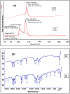
(A) DSC thermogram of mice skin. (A1) Untreated, (A2) Treated with optimized FTQ-NEG. (B) FTIR spectra of mice skin. (B1) Untreated, (B2) Treated with optimized FTQ-NEG.
Figure 4
Figure 4
Confocal laser scanning microscopy in optical sectioning of tissues. (A) pure drug TQ (B) FTQ-NEG normal skin (C) FTQ-NEG with psoriatic skin.
Figure 5
Figure 5
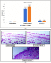
(A) The skin irritation potential score of all the groups. Data are expressed as mean ± SD (*** p < 0.001, control group vs negative control for erythema and edema) and (### p < 0.001, negative control vs FTQ-NEG for erythema and edema). (B) Histopathology of all the treatment groups: Control, FTQ-NEG, and Negative control.
Figure 6
Figure 6
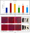
Antipsoriatic experiment in vivo results. Sample picture of balb/c mice from each experimental group: (A) Control group, (B) Negative control, (C) FTQ-NEG, (D) Positive control group, (E) Free drug, and (F) FA loaded in a gel, along with H&E images of histopathological sections from all research groups. Psoriatic plaques in the histological sections are represented with the black arrows.
Figure 7
Figure 7
(A) Evaluation of ear thickness of all the groups. Data are expressed as mean ± SD. Ear thickness was increased in the negative control group in the left ear (** p < 0.01, control group vs negative control for left ear thickness) and decreased in the positive control, FTQ-NEG, FDG, and FA groups compare to the negative control (# p < 0.05), ## p < 0.01 vs negative control) in the left ear and non-significant (ns p > 0.05 vs control) in the right ear. (B) histopathology of the left and right ear of mouse skin of all the groups: Control, Negative control, Positive control, FTQ-NEG, FD, and FA.
Figure 7
Figure 7
(A) Evaluation of ear thickness of all the groups. Data are expressed as mean ± SD. Ear thickness was increased in the negative control group in the left ear (** p < 0.01, control group vs negative control for left ear thickness) and decreased in the positive control, FTQ-NEG, FDG, and FA groups compare to the negative control (# p < 0.05), ## p < 0.01 vs negative control) in the left ear and non-significant (ns p > 0.05 vs control) in the right ear. (B) histopathology of the left and right ear of mouse skin of all the groups: Control, Negative control, Positive control, FTQ-NEG, FD, and FA.
Figure 8
Figure 8
Efficacy study of all the groups. Data are presented as mean ± SD. (A) Body weight was decreased in negative control group when compared to control group (* p < 0.05, control vs negative control on 0th day), (** p < 0.01, control vs negative control on 3rd day & 8th day), (*** p < 0.001, on 5th day) but increased in positive group in comparison to negative control (# p < 0.05). (B) Erythema scoring was significantly increased in negative control group when compared to control group (*** p < 0.001) and decreased in positive control, FTQ-NEG, and FDG groups compare to negative control (# p < 0.05, ## p < 0.01), (### p < 0.001) in erythema before and after. (C) In thickness scoring, negative control group was significantly increased compared to control group (*** p < 0.001) and decreased in positive control, FTQ-NEG, and FDG groups compared to negative control (## p < 0.01) in thickness before and after. (D) In scaling scoring, the negative control group was significantly increased compared to control group (*** p < 0.001) and decreased in positive control, FTQ-NEG, and FDG groups compared to negative control (## p < 0.01) in scaling score after and non-significant in scaling score before.
Figure 9
Figure 9
(A) Data are presented as mean ± SD. Spleen weight decreased in negative control group when compared to control group (*** p < 0.001) but increased in free drug, FTQ-NEG, positive control, and FA groups in comparison to negative control (# p < 0.05, negative control vs free drug), (## p < 0.01, positive control & FA vs negative control),( ### p < 0.001, FTQ-NEG vs negative control). (B) Histopathology of the spleen tissue of all the groups: Control, Negative control, FD, FTQ-NEG, Positive control, and FA.
Figure 10
Figure 10
Immunohistochemistry of skin (1): (A,B) Control group of TNF-α and IL-6, (C,D) Negative control, (E,F) FTQ-NEG, (G,H) FA, (I,J) FD gel, (K,L) Positive control. (2) Immunohistochemistry of spleen: (A,B) Control group of TNF-α and IL-6, (C,D) Negative control group, (E,F) FTQ-NEG group, (G,H) FA, (I,J) FD gel, (K,L) Positive control (IHC, TNF-α, IL-6 400×).
Figure 11
Figure 11
Levels of IL-6 (A) and TNF-α (B) that were assessed in skin homogenate of different animal groups: Control, Negative control, FTQ-NEG, FD, Positive control, and FA (** indicate p < 0.01) and ns is non-significant).

References

    1. Duffin K.C., Chandran V., Gladman D.D., Krueger G.G., Elder J.T., Rahman P. Genetics of Psoriasis and Psoriatic Arthritis: Update and Future Direction. J. Rheumatol. 2008;35:1449–1453. - PMC - PubMed
    1. Griffiths C.E., Barker J.N. Pathogenesis and Clinical Features of Psoriasis. Lancet. 2007;370:263–271. doi: 10.1016/S0140-6736(07)61128-3. - DOI - PubMed
    1. Raychaudhuri S.K., Maverakis E., Raychaudhuri S.P. Diagnosis and Classification of Psoriasis. Autoimmun. Rev. 2014;13:490–495. doi: 10.1016/j.autrev.2014.01.008. - DOI - PubMed
    1. Menter A., Korman N.J., Elmets C.A., Feldman S.R., Gelfand J.M., Gordon K.B., Gottlieb A., Koo J.Y.M., Lebwohl M., Lim H.W., et al. Guidelines of Care for the Management of Psoriasis and Psoriatic Arthritis. J. Am. Acad. Dermatol. 2009;60:4. doi: 10.1016/j.jaad.2008.12.032. - DOI - PubMed
    1. Gupta M.A., Gupta A.K. Depression and Suicidal Ideation in Dermatology Patients with Acne, Alopecia Areata, Atopic Dermatitis and Psoriasis. Br. J. Dermatol. 1998;139:846–850. doi: 10.1046/j.1365-2133.1998.02511.x. - DOI - PubMed

LinkOut - more resources