. 2023 Jan 28;14(2):70.
doi: 10.3390/jfb14020070.

Effect of Sterilization Methods on Electrospun Scaffolds Produced from Blend of Polyurethane with Gelatin

Affiliations

Effect of Sterilization Methods on Electrospun Scaffolds Produced from Blend of Polyurethane with Gelatin

Vera S Chernonosova et al. J Funct Biomater. .

Abstract

Fibrous polyurethane-based scaffolds have proven to be promising materials for the tissue engineering of implanted medical devices. Sterilization of such materials and medical devices is an absolutely essential step toward their medical application. In the presented work, we studied the effects of two sterilization methods (ethylene oxide treatment and electron beam irradiation) on the fibrous scaffolds produced from a polyurethane-gelatin blend. Scaffold structure and properties were studied by scanning electron microscopy (SEM), atomic force microscopy (AFM), infrared spectroscopy (FTIR), a stress-loading test, and a cell viability test with human fibroblasts. Treatment of fibrous polyurethane-based materials with ethylene oxide caused significant changes in their structure (formation of glued-like structures, increase in fiber diameter, and decrease in pore size) and mechanical properties (20% growth of the tensile strength, 30% decline of the maximal elongation). All sterilization procedures did not induce any cytotoxic effects or impede the biocompatibility of scaffolds. The obtained data determined electron beam irradiation to be a recommended sterilization method for electrospun medical devices made from polyurethane-gelatin blends.

Keywords: electron beam irradiation; electrospun scaffold; ethylene oxide; gelatin; polyurethane; protein; sterilization.


Conflict of interest statement

The first two authors should be regarded as joint first authors. Other authors declare no conflict of interest. The funders had no role in the design of the study; in the collection, analyses, or interpretation of data; in the writing of the manuscript; or in the decision to publish the results.

Figures
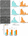
Figure 1
Figure 1
SEM images of scaffolds obtained from a PU-gelatin blend before and after sterilization and distribution of structural parameters (fiber diameter—cyan color diagrams, pore size -orange color diagrams). (A)—unsterilized scaffold; (B)—scaffold sterilized with electron beam irradiation; (C)—scaffold sterilized with ethylene oxide; (D)—statistical analysis of the data.
Figure 2
Figure 2
AFM images of the surface of electrospun scaffolds obtained from a PU-gelatin blend before and after sterilization.
Figure 3
Figure 3
Influence of sterilization on mechanical properties of the scaffolds. (A)—typical tensile stress diagrams of scaffolds before and after sterilization. The plots are presented as the mean of the scaffold strength measured in three samples, bars represent the standard error. (B)—difference in curves for the tensile stress diagrams of materials was obtained using Kolmogorov-Smirnov test.
Figure 4
Figure 4
ATR spectra data or FTIR analysis of the electrospun scaffolds from the PU-gelatin blend (A) and flat films from PU (B) before and after sterilization. Changes in the spectra are marked by vertical dotted lines.
Figure 5
Figure 5
The interaction of fibroblasts with the surfaces of different scaffolds. (A)—Cell viability was tested by Alamar Blue assay after 48-h incubation of cells with scaffolds (Mean ± S.D. of three experiments). (B)—SEM image of cells on the surface of an unsterilized scaffold. (C)—SEM image of cells on the surface of a scaffold sterilized using e-beam irradiation. (D)—SEM image of cells on the surface of a scaffold sterilized using ethylene oxide. SEM images have a magnification of 1000. (E)—Area of cell adhered to the surface materials. The value bars with ns are not significant.

References

    1. Echeverria Molina M.I., Malollari K.G., Komvopoulos K. Design Challenges in Polymeric Scaffolds for Tissue Engineering. Front. Bioeng. Biotechnol. 2021;9:617141. doi: 10.3389/fbioe.2021.617141. - DOI - PMC - PubMed
    1. Perez-Puyana V., Jiménez-Rosado M., Romero A., Guerrero A. Polymer-Based Scaffolds for Soft-Tissue Engineering. Polymers. 2020;12:1566. doi: 10.3390/polym12071566. - DOI - PMC - PubMed
    1. Shoichet M.S. Polymer Scaffolds for Biomaterials Applications. Macromolecules. 2010;43:581–591. doi: 10.1021/ma901530r. - DOI
    1. Ahmad S. Polyurethane: A Versatile Scaffold for Biomedical Applications. SBB. 2018;2:2–4. doi: 10.31031/SBB.2018.02.000536. - DOI
    1. Fathi-Karkan S., Banimohamad-Shotorbani B., Saghati S., Rahbarghazi R., Davaran S. A critical review of fibrous polyurethane-based vascular tissue engineering scaffolds. J. Biol. Eng. 2022;16:6. doi: 10.1186/s13036-022-00286-9. - DOI - PMC - PubMed

LinkOut - more resources