. 2023 Feb 10;14(2):98.
doi: 10.3390/jfb14020098.

Antibacterial Activity and Cell Viability of Biomimetic Magnesian Calcite Coatings on Biodegradable Mg

Affiliations

Antibacterial Activity and Cell Viability of Biomimetic Magnesian Calcite Coatings on Biodegradable Mg

Monica Popa et al. J Funct Biomater. .

Abstract

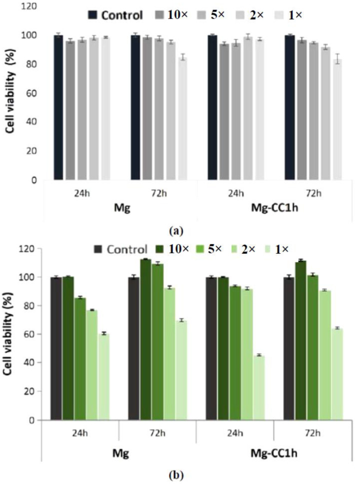
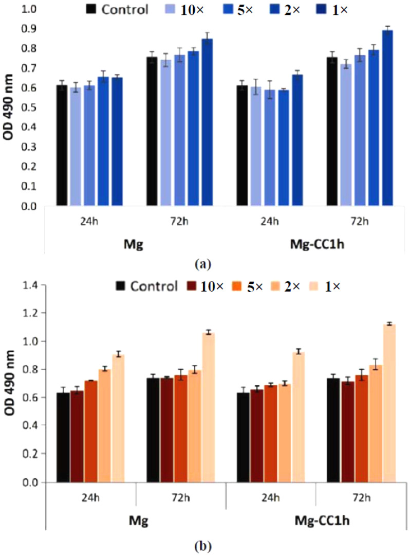
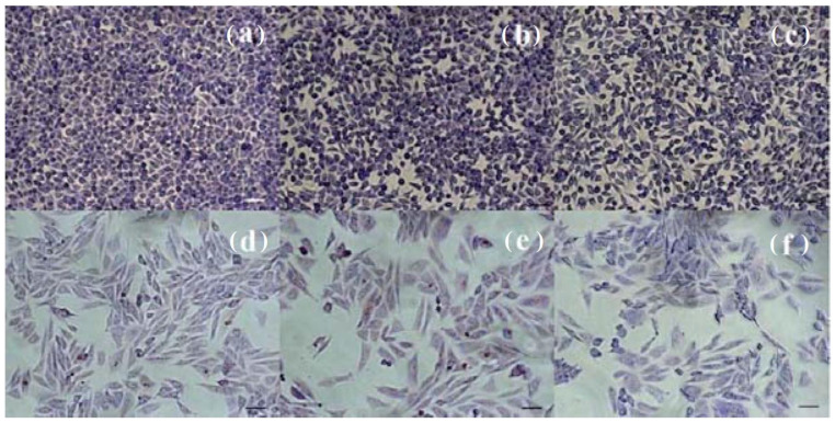
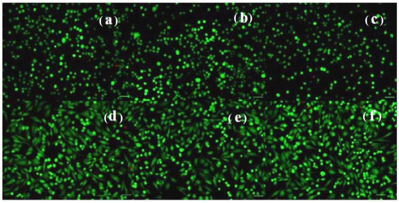
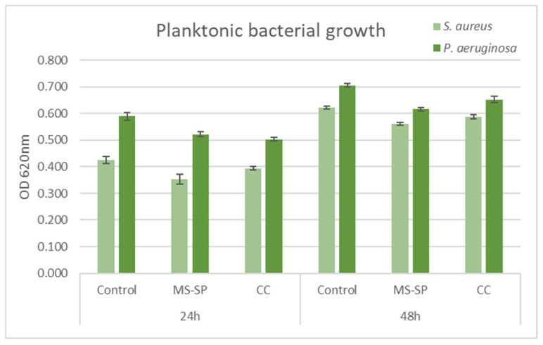
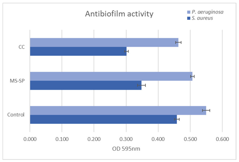
Mg is a material of choice for biodegradable implants. The main challenge for using Mg in temporary implants is to provide protective surfaces that mitigate its rapid degradation in biological fluids and also confer sufficient cytocompatibility and bacterial resistance to Mg-coated surfaces. Even though carbonate mineralization is the most important source of biominerals, such as the skeletons and shells of many marine organisms, there has been little success in the controlled growth of carbonate layers by synthetic processes. We present here the formation mechanism, antibacterial activity, and cell viability of magnesian calcite biomimetic coatings grown on biodegradable Mg via a green, one-step route. Cell compatibility assessment showed cell viability higher than 80% after 72 h using fibroblast cells (NCTC, clone L929) and higher than 60% after 72 h using human osteoblast-like cells (SaOS-2); the cells displayed a normal appearance and a density similar to the control sample. Antimicrobial potential evaluation against both Gram-positive (Staphylococcus aureus (ATCC 25923)) and Gram-negative (Pseudomonas aeruginosa (ATCC 27853)) strains demonstrated that the coated samples significantly inhibited bacterial adhesion and biofilm formation compared to the untreated control. Calcite coatings grown on biodegradable Mg by a single coating process showed the necessary properties of cell compatibility and bacterial resistance for application in surface-modified Mg biomaterials for temporary implants.

Keywords: CaCO3; amorphous calcium carbonate (ACC); antibacterial; bone scaffolds; cell viability; corrosion protective film; resorbable biomaterial.


Conflict of interest statement

The authors declare no conflict of interest. The funders had no role in the design of the study; in the collection, analyses, or interpretation of data; in the writing of the manuscript; or in the decision to publish the results.

Figures

Figure 1
Figure 1
Optical (a) and SEM (b) images of Mg disk after immersion in carbonated water for 30 min, showing the formed CaCO3 coating.
Figure 2
Figure 2
Scheme of surface reactions.
Figure 3
Figure 3
Raman spectra of (a) the Mg surface after 1 min immersion in carbonated water showing the presence of the hydroxyl groups stretching band at 3650 cm−1 and (b) calcite, showing the main carbonate band at 1086 cm−1 and secondary Raman bands of calcite.
Figure 4
Figure 4
Evolution with time of immersion of Raman bands at 3650 cm−1 and 1086 cm−1 representative of the hydroxyl (OH) and carbonate (CO32−) groups.
Figure 5
Figure 5
SEM (a); bi-dimensional (b) and tri-dimensional (c) AFM images showing the surface morphology after 20 min of immersion, scanned on a 1 μm × 1 μm region. Characteristic line scan surface profile (d) showing the nano-scale rugosity of the coating and histogram of the surface nanoparticles size (e) fitted with a Gaussian distribution.
Figure 6
Figure 6
SEM images at different magnifications ((a), 5 µm; (b), 2 µm; (c), 200 nm) showing the nucleation of calcite nano-crystals in the carbonate layer.
Figure 7
Figure 7
Calcite layer formed by the coalescence of growing calcite crystals: SEM ((a), 10 µm; (b), 2 µm; (c), 500 nm).
Figure 8
Figure 8
Scheme illustrating the 2D growth of calcite coating.
Figure 9
Figure 9
SEM (a); bi-dimensional (b) and tri-dimensional (c,d) AFM images taken on a flat region of a crystalline calcite facet; histogram of the surface nanoparticles (e) fitted with a Gaussian distribution; and line profile (f) showing the deposition of nanoparticles on the underlying calcite crystal facet.
Figure 10
Figure 10
Raw bi-dimensional AFM images (unprocessed/as registered) scanned on a 1 μm × 1 μm region on a calcite crystal facet, showing the amplitude (a) and phase contrast (b) after 1 day of immersion. The small particles histogram (c), fitted by a Gaussian distribution, suggests a smaller mean diameter of approximately 11 nm.
Figure 11
Figure 11
Viability of NCTC mouse fibroblasts (a) and SaOS-2 human osteoblasts (b) cultured in the presence of Mg and Mg-CC1h, for 24 h and 72 h, evaluated by the MTT test. The viability of the treated cells was obtained in reference to the negative control (untreated cells), considered to be 100% viable. The data were expressed as the average of the samples analyzed in triplicate (mean ± SD).
Figure 12
Figure 12
The levels of LDH released into the culture medium by NCTC fibroblasts (a) and SaOS-2 human osteoblasts (b) cultured in the presence of Mg and Mg-CC1h for 24 h and 72 h. The data were expressed as the average of the samples analyzed in triplicate (mean ± SD).
Figure 13
Figure 13
Optical microscopy images of NCTC mouse fibroblasts: untreated (a) and treated with dilutions of the Mg (b) and Mg-CC1h samples (c) for 72 h, as well as SaOS-2 osteoblasts: untreated (d) and treated with the dilutions of the Mg (e) and Mg-CC1h samples (f) for 72 h. The untreated cell culture was used as a control (Giemsa staining). Bar = 50 μm.
Figure 14
Figure 14
Fluorescent staining with calcein-AM (green) and ethidium homodimer-1 (red) of NCTC live and dead cells, untreated (a) and treated with solutions of the Mg (b) and Mg-CC1h samples (c) for 24 h; and SaOS-2 osteoblasts untreated (d) and treated with dilutions of the Mg (e) and Mg-CC1h samples (f) for 24 h. Scale bar = 50 μm.
Figure 15
Figure 15
Microbial planktonic growth in the presence of Mg and Mg-CC1h in time (24 and 48 h). Experimental data are reported as mean ± SD. n = 3.
Figure 16
Figure 16
S. aureus and P. aeruginosa biofilm development onto Mg and Mg-CC1h at 72 h. Experimental data are reported as mean ± SD. n = 3.

Similar articles

References

    1. Amini A.R., Laurencin C.T., Nukavarapu S.P. Bone tissue engineering: Recent advances and challenges. Crit. Rev. Biomed. Eng. 2012;40:363–408. doi: 10.1615/CritRevBiomedEng.v40.i5.10. - DOI - PMC - PubMed
    1. Herber V., Okutan B., Antonoglou G., Sommer N.G., Payer M. Bioresorbable magnesium-based alloys as novel biomaterials in oral bone regeneration: General review and clinical perspectives. J. Clin. Med. 2021;10:1842. doi: 10.3390/jcm10091842. - DOI - PMC - PubMed
    1. Song G. Control of biodegradation of biocompatable magnesium alloys. Corros. Sci. 2007;49:1696–1701. doi: 10.1016/j.corsci.2007.01.001. - DOI
    1. Prakasam M., Locs J., Salma-Ancane K., Loca D., Largeteau A., Berzina-Cimdina L. Biodegradable materials and metallic implants—A review. J. Funct. Biomater. 2017;8:44. doi: 10.3390/jfb8040044. - DOI - PMC - PubMed
    1. Hornberger H., Virtanen S., Boccaccini A.R. Biomedical coatings on magnesium alloys—A review. Acta Biomater. 2012;8:2442–2455. doi: 10.1016/j.actbio.2012.04.012. - DOI - PubMed

LinkOut - more resources