

Infrared Macrothermoscopy Patterns-A New Category of Dermoscopy
- PMID: 36826955
- PMCID: PMC9960988
- DOI: 10.3390/jimaging9020036
Infrared Macrothermoscopy Patterns-A New Category of Dermoscopy
Abstract
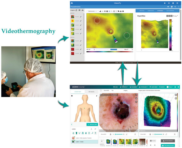
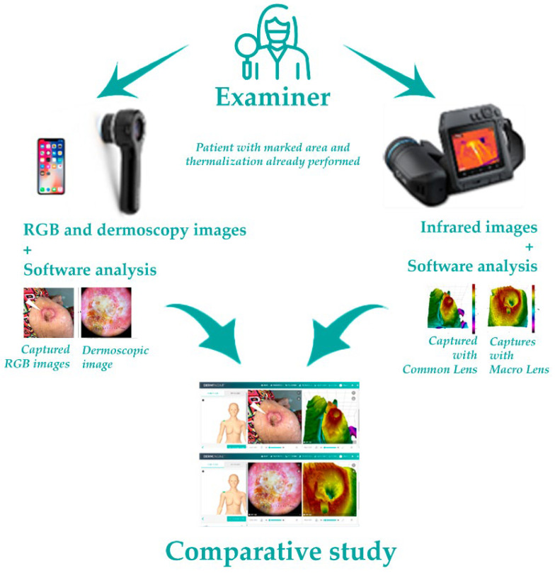
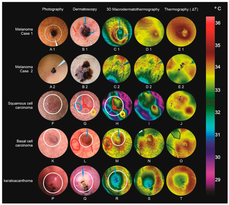
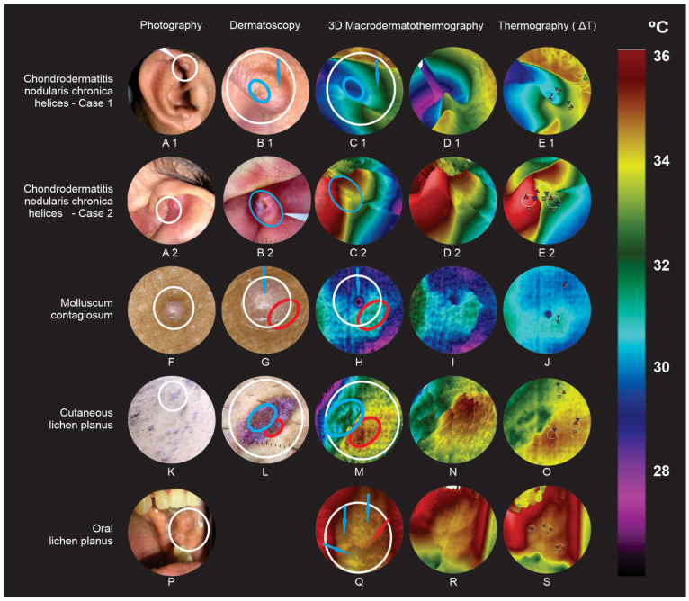
(1) Background: The authors developed a new non-invasive dermatological infrared macroimaging analysis technique (MacroIR) that evaluates microvascular, inflammatory, and metabolic changes that may be dermoscopy complimentary, by analyzing different skin and mucosal lesions in a combined way-naked eye, polarized light dermatoscopy (PLD), and MacroIR-and comparing results; (2) Methods: ten cases were evaluated using a smartphone coupled with a dermatoscope and a macro lens integrated far-infrared transducer into specific software to capture and organize high-resolution images in different electromagnetic spectra, and then analyzed by a dermatologist; (3) Results: It was possible to identify and compare structures found in two dermoscopic forms. Visual anatomical changes were correlated with MacroIR and aided skin surface dermatological analysis, presenting studied area microvascular, inflammatory, and metabolic data. All MacroIR images correlated with PLD, naked eye examination, and histopathological findings; (4) Conclusion: MacroIR and clinic dermatologist concordance rates were comparable for all dermatological conditions in this study. MacroIR imaging is a promising method that can improve dermatological diseases diagnosis. The observations are preliminary and require further evaluation in larger studies.
Keywords: dermatoscopy; dermoscopy; infrared; infrared spectrophotometry; medical thermography; thermography.
Conflict of interest statement
The authors declare no conflict of interest.
Figures
Similar articles
-
Slit lamp polarized dermoscopy: a cost-effective tool to assess eyelid lesions.Int Ophthalmol. 2023 Apr;43(4):1103-1110. doi: 10.1007/s10792-022-02505-0. Epub 2022 Sep 9. Int Ophthalmol. 2023. PMID: 36083562
-
Pre-operative diagnosis of pigmented skin lesions: in vivo dermoscopy performs better than dermoscopy on photographic images.J Eur Acad Dermatol Venereol. 2002 Jul;16(4):339-46. doi: 10.1046/j.1468-3083.2002.00470.x. J Eur Acad Dermatol Venereol. 2002. PMID: 12224689
-
Unusual Clinical Presentation of Giant Extragenital Condyloma.Acta Dermatovenerol Croat. 2020 Dec;28(7):240-241. Acta Dermatovenerol Croat. 2020. PMID: 33834999
-
Ultraviolet-Induced Fluorescence Dermoscopy, a Novel Diagnostic Technique in Dermatological Practice: A Systematic Review.Indian Dermatol Online J. 2024 Dec 13;16(1):25-39. doi: 10.4103/idoj.idoj_299_24. eCollection 2025 Jan-Feb. Indian Dermatol Online J. 2024. PMID: 39850698 Free PMC article. Review.
-
Role of In Vivo Reflectance Confocal Microscopy in the Analysis of Melanocytic Lesions.Acta Dermatovenerol Croat. 2018 Apr;26(1):64-67. Acta Dermatovenerol Croat. 2018. PMID: 29782304 Review.
Cited by
-
Advancements in Dermatological Imaging Modalities.Indian Dermatol Online J. 2024 Feb 28;15(2):278-292. doi: 10.4103/idoj.idoj_852_23. eCollection 2024 Mar-Apr. Indian Dermatol Online J. 2024. PMID: 38550821 Free PMC article. No abstract available.
References
-
- Phillips M., Marsden H., Jaffe W., Matin R.N., Wali G.N., Greenhalgh J., McGrath E., James R., Ladoyanni E., Bewley A., et al. Assessment of Accuracy of an Artificial Intelligence Algorithm to Detect Melanoma in Images of Skin Lesions. JAMA Netw. Open. 2019;2:e1913436. doi: 10.1001/jamanetworkopen.2019.13436. - DOI - PMC - PubMed
LinkOut - more resources
Full Text Sources
