. 2023 Jan 19;21(2):61.
doi: 10.3390/md21020061.

Characterization of the Native Disulfide Isomers of the Novel χ-Conotoxin PnID: Implications for Further Increasing Conotoxin Diversity

Affiliations

Characterization of the Native Disulfide Isomers of the Novel χ-Conotoxin PnID: Implications for Further Increasing Conotoxin Diversity

Michael J Espiritu et al. Mar Drugs. .

Abstract

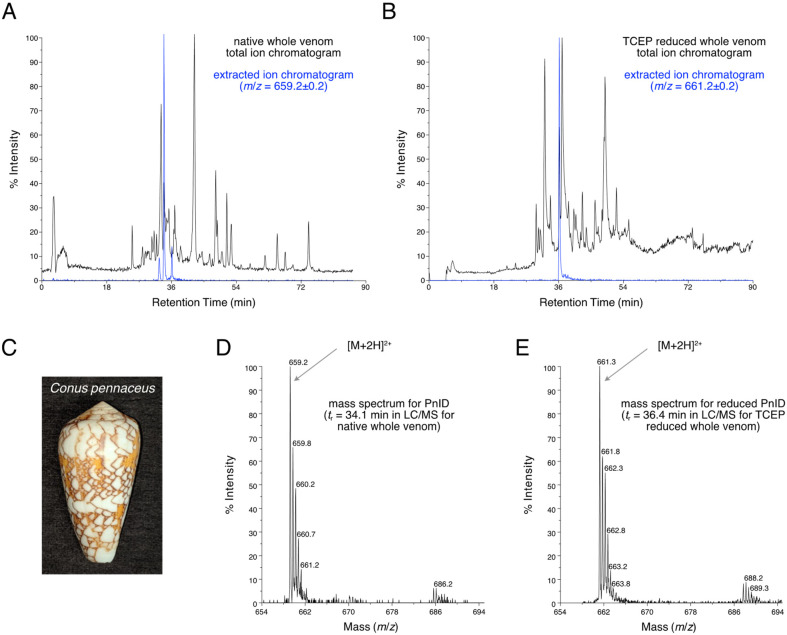
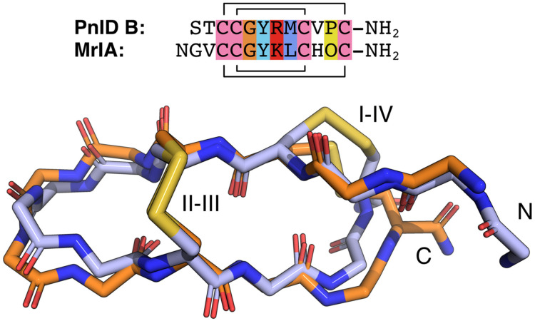
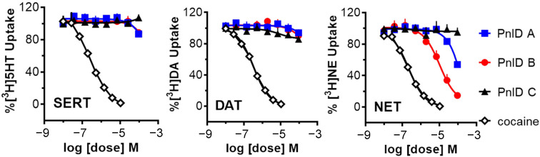
χ-Conotoxins are known for their ability to selectively inhibit norepinephrine transporters, an ability that makes them potential leads for treating various neurological disorders, including neuropathic pain. PnID, a peptide isolated from the venom of Conus pennaceus, shares high sequence homology with previously characterized χ-conotoxins. Whereas previously reported χ-conotoxins seem to only have a single native disulfide bonding pattern, PnID has three native isomers due to the formation of different disulfide bond patterns during its maturation in the venom duct. In this study, the disulfide connectivity and three-dimensional structure of these disulfide isomers were explored using regioselective synthesis, chromatographic coelution, and solution-state nuclear magnetic resonance spectroscopy. Of the native isomers, only the isomer with a ribbon disulfide configuration showed pharmacological activity similar to other χ-conotoxins. This isomer inhibited the rat norepinephrine transporter (IC50 = 10 ± 2 µM) and has the most structural similarity to previously characterized χ-conotoxins. In contrast, the globular isoform of PnID showed more than ten times less activity against this transporter and the beaded isoform did not display any measurable biological activity. This study is the first report of the pharmacological and structural characterization of an χ-conotoxin from a species other than Conus marmoreus and is the first report of the existence of natively-formed conotoxin isomers.

Keywords: conotoxins; isomers; monoamine transporters; peptide; structure; toxins.


Conflict of interest statement

The authors declare no conflict of interest. The funders had no role in the design of the study; in the collection, analyses, or interpretation of data; in the writing of the manuscript; or in the decision to publish the result.

Figures

Figure 1
Figure 1
(A). LC-MS total ion chromatogram of the crude duct venom extract of Red Sea C. pennaceus (pictured in (C)). The extracted ion chromatogram for m/z = 659.2 (shown in blue) has peaks corresponding to the [M+2H]2+ ions of the native PnID peptides. (B). The total ion chromatogram LC-MS after TCEP reduction. The extracted ion chromatogram for m/z = 661.2 (shown in blue) has only a single peak, corresponding to the [M+2H]2+ ion of fully reduced PnID. (D). Mass spectrum corresponding to a retention time of 34.1 min in the LC-MS data shown in (A). has an m/z value consistent with the expected value for the [M+2H]2+ ion of fully oxidized PnID. (E). Mass spectrum corresponding to a retention time of 36.4 min in the LC-MS data shown in (B). has an m/z value consistent with the expected value for the fully-reduced [M+2H]2+ ion of PnID.
Figure 2
Figure 2
Comparative RP-HPLC analysis of the single-step (random) oxidized isomers of PnID to the regioselectively synthesized forms. Numbers above each sequence indicate the positions of the cysteines. (A). The isomeric products of the random air oxidation of PnID. (BD). Co-elution of the selectively folded material with the random air oxidation products. * Indicates position of the mixed peak, the arrow indicates the appearance of shouldering and a lack of coelution. (B,C). are the same scale, (A,D). are rescaled to fit.
Figure 3
Figure 3
Ensemble (top) and cartoon (bottom) representations of PnID A (left) and PnID B (right). The 20 lowest-energy structures from 100 structures calculated in the final iteration of the ARIA protocol were refined in explicit water and used to generate the structural ensembles. The N-termini are colored blue, the C-termini are colored red, and the disulfide bond topologies are indicated by Roman numerals. The same orientation and scale are used for the ensemble (top) and cartoon (bottom) representations. The cartoon representations show the lowest energy structure from each ensemble. For purposes of clarity, only 10 of the 20 structures used to define the structural ensemble are shown here with residues 2–11 aligned.
Figure 4
Figure 4
Sequence and backbone alignments for PnID B (orange) and MrIA (blue). The backbone atoms for the lowest energy structure for the PnID B structural ensemble (in orange) were aligned against the backbone atoms for the lowest energy structure (in blue) from the ensemble for MrIA (PDB ID 2EW4). The sequence alignment for these peptides is provided above and colored following the ClustalX default coloring scheme. In the MrIA sequence, “O” represents hydroxyproline. Both peptides have an amidated C-terminus (NH2).
Figure 5
Figure 5
Uptake inhibition plots showing the comparative dose-response inhibition of the monoamine transporters SERT (left), DAT (middle), and NET (right) by PnID A, PnID B, PnID C, and cocaine. Whereas the PnID isomers do not show inhibition at SERT and DAT, the ribbon isoform (PnID B) shows activity for inhibiting NET. Statistical analyses were carried out using GraphPad Prism 6.0 (GraphPad Scientific, San Diego, CA, USA). IC50 values for uptake inhibition were calculated based on non-linear regression analysis.

References

    1. Kaas Q., Westermann J.-C., Halai R., Wang C.K.L., Craik D.J. ConoServer, a Database for Conopeptide Sequences and Structures. Bioinformatics. 2008;24:445–446. doi: 10.1093/bioinformatics/btm596. - DOI - PubMed
    1. Kaas Q., Yu R., Jin A.-H., Dutertre S., Craik D.J. ConoServer: Updated Content, Knowledge, and Discovery Tools in the Conopeptide Database. Nucleic Acids Res. 2012;40:D325–D330. doi: 10.1093/nar/gkr886. - DOI - PMC - PubMed
    1. Jin A.-H., Muttenthaler M., Dutertre S., Himaya S.W.A., Kaas Q., Craik D.J., Lewis R.J., Alewood P.F. Conotoxins: Chemistry and Biology. Chem. Rev. 2019;119:11510–11549. doi: 10.1021/acs.chemrev.9b00207. - DOI - PubMed
    1. Lavergne V., Harliwong I., Jones A., Miller D., Taft R.J., Alewood P.F. Optimized Deep-Targeted Proteotranscriptomic Profiling Reveals Unexplored Conus Toxin Diversity and Novel Cysteine Frameworks. Proc. Natl. Acad. Sci. USA. 2015;112:E3782–E3791. doi: 10.1073/pnas.1501334112. - DOI - PMC - PubMed
    1. Zhou J. Norepinephrine Transporter Inhibitors and Their Therapeutic Potential. Drugs Future. 2004;29:1235–1244. doi: 10.1358/dof.2004.029.12.855246. - DOI - PMC - PubMed

LinkOut - more resources