

Is retina affected in Huntington's disease? Is optical coherence tomography a good biomarker?
- PMID: 36827300
- PMCID: PMC9955964
- DOI: 10.1371/journal.pone.0282175
Is retina affected in Huntington's disease? Is optical coherence tomography a good biomarker?
Abstract
Aim of the study: Comparative cross-sectional study of retinal parameters in Huntington's disease and their evaluation as marker of disease progression.
Clinical rationale for the study: Huntington's disease (HD) is a neurodegenerative disorder with dominant motor and neuropsychiatric symptoms. Involvement of sensory functions in HD has been investigated, however studies of retinal pathology are incongruent. Effect sizes of previous findings were not published. OCT data of the subjects in previous studies have not been published. Additional examination of structural and functional parameters of retina in larger sample of patients with HD is warranted.
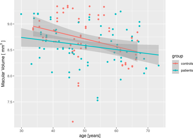
Materials and methods: This is a prospective cross-sectional study that included: peripapillary retinal nerve fiber layer thickness (RNFL) and total macular volume (TMV) measured by spectral domain optical coherence tomography (OCT) of retina, Pelli-Robson Contrast Sensitivity test, Farnsworth 15 Hue Color discrimination test, ophthalmology examination and Unified Huntington's disease Rating Scale (UHDRS). Ninety-four eyes of 41 HD patients examined in total 47 visits and 82 eyes of 41 healthy controls (HC) examined in total 41 visits were included. Analyses were performed by repeated measures linear mixed effects model with age and gender as covariates. False discovery rate was corrected by Benjamini-Hochberg procedure.
Results: HD group included 21 males and 20 females (age 50.6±12.0 years [mean ± standard deviation], disease duration 7.1±3.6 years, CAG triplet repeats 44.1±2.4). UHDRS Total Motor Score (TMS) was 30.0±12.3 and Total Functional Capacity 8.2±3.2. Control group (HC) included 19 males and 22 females with age 48.2±10.3 years. There was no statistically significant difference between HD and HC in age. The effect of the disease was not significant in temporal segment RNFL thickness. It was significant in the mean RNFL thickness and TMV, however not passing false discovery rate adjustment and with small effect size. In the HD group, the effect of disease duration and TMS was not significant. The Contrast Sensitivity test in HD was within normal limits and the 15-hue-test in HD did not reveal any specific pathology.
Conclusions: The results of our study support possible diffuse retinal changes in global RNFL layer and in macula in Huntington's disease, however, these changes are small and not suitable as a biomarker for disease progression. We found no other structural or functional changes in retina of Huntington's disease patients using RNFL layer and macular volume spectral domain OCT and Contrast Sensitivity Test and 15-hue-test.
Clinical implications: Current retinal parameters are not appropriate for monitoring HD disease progression.
Copyright: © 2023 Dusek et al. This is an open access article distributed under the terms of the Creative Commons Attribution License, which permits unrestricted use, distribution, and reproduction in any medium, provided the original author and source are credited.
Conflict of interest statement
The authors have declared that no competing interests exist.
Figures
References
-
- Johnson M, Gelderblom H, Rüther K, Priller J, Bernstein SL. Evidence that Huntington’s disease affects retinal structure and function. Investigative Ophtalmology & Visual Science 2014; Vol. 55, 1644.
Publication types
MeSH terms
Substances
LinkOut - more resources
Full Text Sources
Other Literature Sources
Medical
Research Materials
