. 2023 Feb 24;9(8):eade8653.
doi: 10.1126/sciadv.ade8653. Epub 2023 Feb 24.

A microbiota and dietary metabolite integrates DNA repair and cell death to regulate embryo viability and aneuploidy during aging

Affiliations

A microbiota and dietary metabolite integrates DNA repair and cell death to regulate embryo viability and aneuploidy during aging

Robert Sonowal et al. Sci Adv. .

Abstract

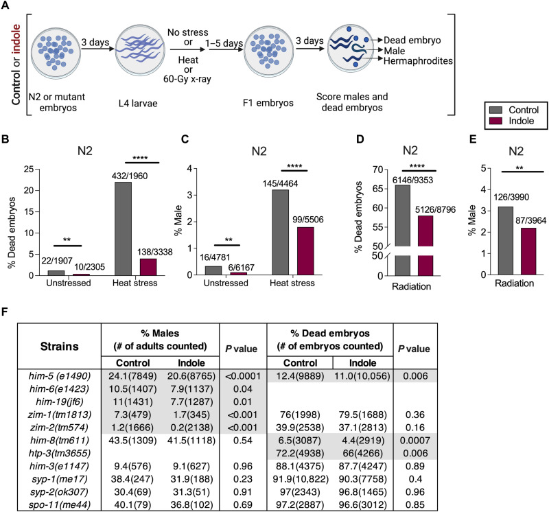
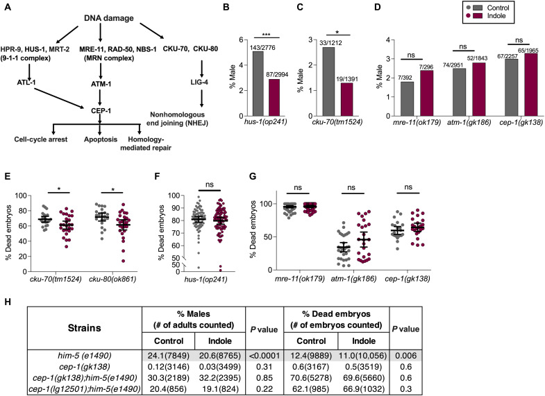
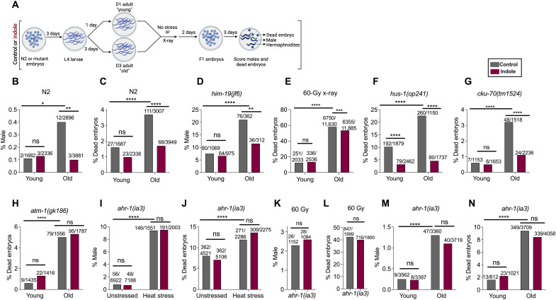
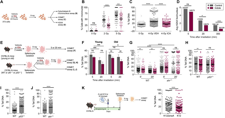
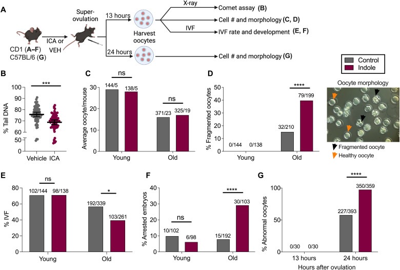
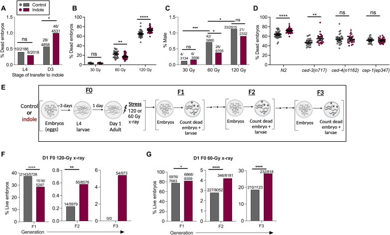
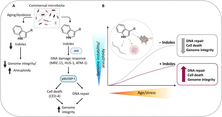
During aging, environmental stressors and mutations along with reduced DNA repair cause germ cell aneuploidy and genome instability, which limits fertility and embryo development. Benevolent commensal microbiota and dietary plants secrete indoles, which improve healthspan and reproductive success, suggesting regulation of germ cell quality. We show that indoles prevent aneuploidy and promote DNA repair and embryo viability, which depends on age and genotoxic stress levels and affects embryo quality across generations. In young animals or with low doses of radiation, indoles promote DNA repair and embryo viability; however, in older animals or with high doses of radiation, indoles promote death of the embryo. These studies reveal a previously unknown quality control mechanism by which indole integrates DNA repair and cell death responses to preclude germ cell aneuploidy and ensure transgenerational genome integrity. Such regulation affects healthy aging, reproductive senescence, cancer, and the evolution of genetic diversity in invertebrates and vertebrates.


Figures

Fig. 1.
Fig. 1.. Indoles limit intergenerational embryo lethality and male frequency induced by heat stress, X-irradiation, and mutations in C. elegans.
(A) Schematic for assessing male frequency and embryo lethality in the progeny of N2 or mutant worms grown either under control or under indole conditions. Indole or carrier was provided throughout the experiment. (B and D) Percentage of dead embryos produced by N2, subjected to either 30°C (B) or 60 Gy (D). (C and E) Frequency of males in the progeny of N2 exposed to either 30°C (C) or 60 Gy (E). Values on bars in (B) and (D) represent the number of dead embryos/total embryos counted, and bars in (C) and (E) represent the number of males/total live progeny counted. (F) Frequency of males and dead embryos produced by mutant worms. P values calculated by chi-square test. Combined results from at least two independent experiments are presented. **P < 0.01and ****P < 0.0001.
Fig. 2.
Fig. 2.. Indoles require the MRN pathway genes to limit male frequency and embryo lethality in C. elegans.
(A) Schematic of DNA damage response (DDR) pathways in C. elegans. In C. elegans germline, DNA DSBs are detected by either the HPR-9-HUS-1-MRT-2 (9-1-1) sensor complex or by the MRN sensor complex, which activate the C. elegans p53 homolog CEP-1. An alternative DSB sensor induces nonhomologous end joining (NHEJ) in somatic cells, although this pathway is suppressed within the gonad (64). The MRN-p53/CEP-1 pathway regulates homology-mediated DSB repair during meiosis, and following genotoxic stress (39), p53/CEP-1 induces apoptosis via apoptosis-activating factor-1 (APAF-1) homolog and a caspase (20, 65). (B to G) DDR mutant worms were grown in either control or indole, exposed to 60-Gy x-ray. Indole or carrier was provided throughout the experiments. (B to D) Frequencies of males in the progeny (values on bars represent total number of males/total live progeny counted). (E to G) Viability of embryos produced (n > 25 biological replicates/condition). (H) Male frequency, and embryo lethality in the progeny of worms harboring DDR mutations, and grown either under control or under indole conditions. Indole or carrier was provided throughout the experiments. P values in (B) to (D) and (H) calculated by chi-square test. Values in (E) to (G) represent mean values ±95% CI, and P values were calculated by Mann-Whitney test. Combined results from at least two independent experiments are presented. *P < 0.05 and ***P < 0.001. ns, not significant.
Fig. 3.
Fig. 3.. Indoles limit incidence of male and embryo lethality associated with stressor or aging in C. elegans via MRN and Ahr-1 pathways.
(A) Schematic for assessing male frequency and embryo lethality induced with aging or stressors in C. elegans. Worms were grown either under control or under indole conditions for the duration of all experiments. Frequencies of male or dead embryos in the progeny of young [day 1 (D1) to day 3 (D3) of adulthood] and old (D3 to D5 of adulthood) N2 (B and C) or him-19 mutant animals (D). (E) Percentage of dead embryos in the progeny from young and old N2 worms, subjected to 60 Gy, and allowed to lay embryos for 48 hours. (F to H) Frequencies of dead embryos in the progeny of young and old DDR mutant worms. Frequency of males (I and K) and dead embryos (J and L) in the progeny of ahr-1(ia3) worms, subjected to either 30°C heat stress (I and J) or 60 Gy (K and L). Frequencies of males (M) and dead embryos (N) in the progeny of young and old ahr-1(ia3) worms. Values on the top of the bars represent total number of males/total live progeny counted in (B), (D), (I), (K), and (M), and number of dead embryos/total embryos counted in (C), (E) to (H), (J), (L), and (N). Combined results from at least two independent experiments are presented. P values calculated by chi-square test. *P < 0.05, **P < 0.01, ***P < 0.001, and ****P < 0.0001.
Fig. 4.
Fig. 4.. Indoles limit x-ray–induced DNA damage and promote DNA repair in fibroblasts and splenocytes.
(A) Schematic of DDR assays. 3T3 fibroblast cells were treated with vehicle or ICA and assayed for the frequency of micronuclei with different radiation doses (B), percent comet tail DNA (n > 102 comets per condition; C), and H2AX foci after 4-Gy x-ray (D). Values above the bars represent number of foci observed/total nuclei counted. (E) Schematic of DDR assays in splenocytes from C57BL/6 mice. (F to K) The percentage (%) of tail DNA in comets. (F) Four-gray irradiated splenocytes isolated from ICA/vehicle-treated young (3-month) and old (18-month) C57BL/6 mice (n > 102 comets per condition). (G) Four-gray irradiated splenocytes from ahr−/− mice (n > 200 comets per condition; comet assay performed 5′ and 20′ after radiation). (H) Four-gray irradiated splenocytes from p53−/− mice (n > 170 comets per condition). (I) Splenocytes from p53−/− mice without stress (n > 230 comets per condition). (J) Splenocytes from ahr−/− (n > 360 comets per condition) and age-matched C57BL/6 mice without stress. (K) Four-gray irradiated splenocytes from C57BL/6 mice colonized with either K12 or K12∆tnaA (n > 200 comets per condition). Values represent combined results from at least two independent experiments. (B), (C), and (F) to (K) represent mean values ±95% CI. P values calculated with Mann-Whitney test (B, C, and G to K), Kruskal-Wallis (F) or chi-square test (D). *P < 0.05, **P < 0.01, ***P < 0.001, and ****P < 0.0001.
Fig. 5.
Fig. 5.. Indoles regulate DNA repair and survival of mouse oocytes.
(A) Schematic of mouse oocyte quality assessment. (B) Percentage of tail DNA in the comets of X-irradiated CD1 oocytes from mice treated with ICA or vehicle (n > 70 comets per condition). (C to F) Quality assessment of the oocytes from young (3 months old) or old (> 6 months) CD1 mice treated with ICA or vehicle. (C) Oocyte number per mouse. Values above the bars represent the number of oocytes/total number mice. (D) Frequency of fragmented oocytes. Values above the bars represent the number of fragmented oocytes/total oocytes. The right panel in (D) represents morphologies of normal versus fragmented (abnormal) oocytes (magnification, ×40). (E) The IVF success rate. Values above the bars represent the number of fertilized oocytes/total number of oocytes. (F) Frequency of developmental arrest observed in embryos after IVF. Values above the bars represent the number of arrested embryos/total embryos scored. (G) Frequency of abnormal oocytes from young (3 months old) C57BL/6 mice ICA/vehicle treated for 2 days and subjected to post-ovulatory aging. Values above the bars represent number of abnormal oocytes/total number of oocytes. Data represent combined results from at least two independent experiments. In (B), data represent mean values ±95% CI. P values were calculated with Mann-Whitney test (B) or chi-square test (C to G). *P < 0.05, ***P < 0.001, and ****P < 0.0001.
Fig. 6.
Fig. 6.. Indoles regulate DNA repair and cell survival depending on age and level of damage to promote transgenerational survival in C. elegans.
(A) Frequency of dead embryos from N2 grown under control conditions and transferred to indole condition either at the L4 or D3 stage (values represent “number of dead embryos/total number of embryos”). (B) Frequency of dead embryos. (C) Male progeny from control/vehicle grown D1 adult N2, subjected to 30-, 60-, and 120-Gy x-ray [values in (C) represent “number of males/total number of adults scored”]. (D) Frequency of dead embryos from D1 N2 or cep-1, ced-3, and ced-4 mutants subjected to 120-Gy x-ray. (E) Schematic of transgenerational embryo survival assays. (F and G) Frequency of live embryos obtained until F3 generation when F0 D1 N2 adults grown either in control or in indole were exposed to either 120 Gy (F) or 60 Gy (G) x-ray (values represent “number of live embryos/total number of embryos scored”). All values represent combined results from at least two independent experiments. Data in (B) and (D) represent mean values ±95% CI. P values were calculated with Mann-Whitney test (B and D) or chi-square test (A, C, F, and G). *P < 0.05, **P < 0.01, ***P < 0.001, and ****P < 0.0001
Fig. 7.
Fig. 7.. Model of indole quality control regulation.
(A) Indole and the indole derivative ICA are derived from the microbiota or from dietary sources (e.g., cruciferous vegetables) and act via AhR, MRN-1, and p53/CEP-1 to detect DNA damage and activate the DSB repair machinery when damage is reparable, and cell death via the apoptosis activating factor CED4 when it is not, ensuring genome integrity and homeostasis. Alternatively, dysbiosis, dietary changes, or aging results in decreased levels of indole and thereby increased aneuploidy and genome variability. (B) Indoles act in a “quality control” capacity via MRN and p53 to affect genome integrity. In youth or low stressor levels, repair capacity is high, and indole has little effect. With age or increasing stressor levels, indole augments repair together with cell death, so that surviving cells have higher genomic integrity but more limited diversity compared to cells without indole. Thus, cell death removes heavily damaged cells, limiting their impact on diversity and fecundity. Increasing either repair or cell death increases the percentage of F1 animals with intact genomes, providing a survival advantage in successive generations. Limiting indole, which occurs with dysbiosis, dietary changes, or aging, relaxes the p53 checkpoint, resulting in decreased repair and cell death, increased aneuploidy, and, in worms, more males. Males outcross via sexual reproduction, increasing genomic diversity and allowing rapid adaptation to changing environments. Worm and mammalian oocytes have different set points for death and repair, with worms favoring survival and repair over death and mammals favoring the opposite. We speculate that species differences arise from selection for intact genomes when sequentially generating small numbers of highly complex live young, which is metabolically expensive, versus generating large numbers of embryos.

References

    1. T. E. Schmid, B. Eskenazi, A. Baumgartner, F. Marchetti, S. Young, R. Weldon, D. Anderson, A. J. Wyrobek, The effects of male age on sperm DNA damage in healthy non-smokers. Hum. Reprod. 22, 180–187 (2007). - PubMed
    1. K. Selesniemi, H. J. Lee, A. Muhlhauser, J. L. Tilly, Prevention of maternal aging-associated oocyte aneuploidy and meiotic spindle defects in mice by dietary and genetic strategies. Proc. Natl. Acad. Sci. U.S.A. 108, 12319–12324 (2011). - PMC - PubMed
    1. J. Carroll, P. Marangos, The DNA damage response in mammalian oocytes. Front. Genet. 4, 117 (2013). - PMC - PubMed
    1. P. Nurse, Y. Masui, L. Hartwell, Understanding the cell cycle. Nat. Med. 4, 1103–1106 (1998). - PubMed
    1. L. Stergiou, M. O. Hengartner, Death and more: DNA damage response pathways in the nematode C. elegans. Cell Death Differ. 11, 21–28 (2004). - PubMed