. 2023 Feb 24;18(2):e0279655.
doi: 10.1371/journal.pone.0279655. eCollection 2023.

High glucose induces an activated state of partial epithelial-mesenchymal transition in human primary tubular cell cultures

Affiliations

High glucose induces an activated state of partial epithelial-mesenchymal transition in human primary tubular cell cultures

Barbara Torsello et al. PLoS One. .

Abstract

Tubulointerstitial fibrosis is observed in diabetic nephropathy. It is still debated whether tubular cells, undergoing epithelial-mesenchymal transition (EMT) in high glucose (HG) conditions, may contribute to interstitial fibrosis development. In this study, we investigated the phenotypic and molecular EMT-like changes and the alteration of inflammatory and fibrogenic secretome induced by HG in human primary tubular cell cultures. Taking advantage of this in vitro cell model composed of proximal and distal tubular cells, we showed that HG-treated tubular cells acquired a fibroblast-like morphology with increased cytoplasmic stress fibers, maintaining the expression of the epithelial markers specific of proximal and distal tubular cells. HG increased Snail1, miRNA210 and Vimentin mesenchymal markers, decreased N-cadherin expression and migration ability of primary tubular cells, while E-cadherin expression and focal adhesion distribution were not affected. Furthermore, HG treatment of tubular cells altered the inflammatory cytokine secretion creating a secretome able to enhance the proliferation and migration of fibroblasts. Our findings show that HG promotes an activated state of partial EMT in human tubular primary cells and induces a pro-inflammatory and pro-fibrogenic microenvironment, supporting the active role of tubular cells in diabetic nephropathy onset.


Conflict of interest statement

The authors have declared that no competing interests exist.

Figures
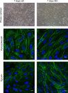
Fig 1
Fig 1. Representative morphological evaluation and cell distribution of epithelial markers in primary tubular cell cultures grown for 7 days in control (ctrl) or high glucose (HG) medium.
The phase contrast images (200x) show the different cell morphology. The confocal microscopy images show the cell distribution of the epithelial markers Pancytokeratin and Epcam (green). DAPI (blue) was used to counterstain the nuclei. Scale bars: 10 μm.
Fig 2
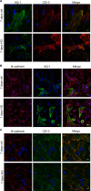
Fig 2. Cell distribution of proximal tubular markers in primary tubular cell cultures grown for 7 days in control (ctrl) or high glucose (HG) medium.
Representative confocal microscopy images of cell cultures stained with antibodies against: (A) Aquaporin-1 (AQ-1, green) and CD13 (red); (B) N-cadherin (red) and AQ-1 (green); (C) N-cadherin (red) and CD13 (green). The merge panels show the co-localization signals. DAPI (blue) was used to counterstain the nuclei. Scale bars: 10 μm.
Fig 3
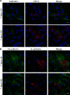
Fig 3. Cell distribution of proximal and distal tubular markers in primary tubular cell cultures grown for 7 days in control (ctrl) or high glucose (HG) medium.
Representative confocal microscopy images of cell cultures stained with antibodies against: (A) Calbindin (green) and CD13 (red); (B) N-cadherin (green) and E-cadherin (red). The merge panels show the co-localization signals. DAPI (blue) was used to counterstain the nuclei. Scale bars: 10 μm.
Fig 4
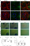
Fig 4. Stress fibers and focal adhesion cellular distribution and migration evaluated in primary tubular cell cultures grown for 96h in control (ctrl) or high glucose (HG) medium.
(A) Representative confocal microscopy images of cell cultures stained with Alexa-Fluor-594-phalloidin (red) and anti-Paxillin antibody (green). DAPI (blue) was used to counterstain the nuclei. Scale bars: 10 μm. (B) Representative phase contrast images of wound healing assay at the scratch (0h) and after 8h of wound recovering performed in primary tubular cultures grown for 96h in control (ctrl) and high glucose (HG) medium. Scale bars: 100 μm. (C) Representative images of Haematoxylin-Eosin stained tubular cells grown in control (ctrl) and high glucose (HG) medium for 96h and migrated in Boyden chamber for 6h. (D) Wound healing graph representing the recovery expressed as μm in three independent experiments. (E) Boyden assay graph reporting the mean number of migrating cells counted in 10 randomly chosen fields for each sample in four independent experiments. *p<0.05.
Fig 5
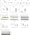
Fig 5. EMT marker expression in primary tubular cell cultures grown in control (ctrl) and high glucose (HG) medium.
(A) Snail1, Zeb2, E-cadherin, Col1a2, and Fibronectin gene expression and (B) miRNA210, miRNA145 and miRNA149 expression evaluated by Real-time PCR at the indicated time points in control (ctrl) and high glucose (HG) conditions. The relative amounts of the different transcripts and miRNAs, calculated as 2−ΔΔCt, are expressed as fold change with respect to the corresponding control sample equal to 1. (n≥3). *p<0.05. (C) Representative western blot of Vimentin, N-cadherin, E-cadherin and β-actin proteins evaluated in lysates of primary tubular cell cultures grown for the indicated time points in control (ctrl) and high glucose (HG) medium. In the box plots the densitometric values of the specific protein bands normalised for corresponding β-actin bands are expressed as a fold change with respect to corresponding control samples. (n≥4). *p<0.05. (D) Representative western blot of α-SMA protein evaluated in lysates of primary tubular cell cultures grown for the indicated time points in control (ctrl) and high glucose (HG) medium. The lysate of LX-2 human fibroblast cell line is used as α-SMA positive control.
Fig 6
Fig 6. Cytokines, chemokines and growth factors differently secreted by primary tubular cell cultures grown for the indicated time points in control (ctrl) and high glucose (HG) medium.
Cytokines increased (A) or decreased (B) in HG conditioned medium of primary tubular cell cultures compared to control. The plots show the mean concentration of the cytokines, expressed as pg/mg, in control conditioned medium (white circles; ctrl) and HG conditioned medium (white triangles; HG) of two independent experiments analyzed in duplicate. *p<0.05.
Fig 7
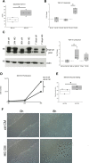
Fig 7. Evaluation of TGF-β1 secretion and expression after HG treatment of primary tubular cell cultures, and effects of conditioned media of HG-treated tubular cells on murine fibroblast proliferation and migration.
(A) Quantification by ELISA of TGF-β1 secreted in conditioned medium of tubular cells cultured for 96h in control (ctrl) and high glucose (HG) medium. Data are expressed as pg/mg in four independent experiments analysed in duplicate. *p<0.05. (B) TGF-β1 transcript expression evaluated by Real-time PCR in primary tubular cell cultures at the indicated time points in control (ctrl) and high glucose (HG) conditions. The relative transcript amounts, calculated as 2−ΔΔCt, are represented as fold change with respect to the corresponding control sample considered equal to 1. (n = 3). *p<0.05. (C) Representative western blot of TGF-β1 precursor and β-actin proteins evaluated in lysates of primary tubular cell cultures grown for the indicated time points in control (ctrl) and high glucose (HG) medium. In the box plots, the densitometric values of the TGF-β1 precursor bands normalised for corresponding β-actin bands are expressed as fold change with respect to corresponding control samples. (n≥3). *p<0.05. (D) Growth curves of NIH3T3 fibroblasts cultured in control conditioned medium (white squares, ctrl CM) and in high glucose conditioned medium (white triangles, HG CM) of primary tubular cell cultures. (n = 3). *p<0.05. (E) Wound healing graph representing the recovery of NIH3T3 fibroblasts treated with conditioned medium of primary tubular cell cultures grown for 96h in control (ctrl CM) and HG (HG CM) medium. Data are expressed as μm in three independent experiments. *p<0.05. (F) Representative phase contrast images of wound healing assay at the scratch (0h) and after 8h of wound recovering performed in NIH3T3 fibroblasts treated with conditioned medium of primary tubular cultures grown for 96h in control (ctrl CM) and high glucose (HG CM) medium.

References

    1. Rao VR, Tan SH, Candasamy M, Bhattamisra SK. Diabetic nephropathy: An update on pathogenesis and drug development. Diabetes Metab Syndr Clin Res Rev. 2019; 13: 754–762. doi: 10.1016/j.dsx.2018.11.054 - DOI - PubMed
    1. Garcia-Fernandez N, Jacobs-Cachá C, Mora-Gutiérrez JM, Vergara A, Orbe J, Soler MJ. Matrix metalloproteinases in diabetic kidney disease. J Clin Med. 2020; 9: 1–17. doi: 10.3390/jcm9020472 - DOI - PMC - PubMed
    1. Lee YJ, Han HJ. Troglitazone ameliorates high glucose-induced EMT and dysfunction of SGLTs through PI3K/Akt, GSK-3β, Snail1, and β-catenin in renal proximal tubule cells. Am J Physiol—Ren Physiol. 2010; 298: 1263–1275. doi: 10.1152/ajprenal.00475.2009 - DOI - PubMed
    1. Hills CE, Siamantouras E, Smith SW, Cockwell P, Liu KK, Squires PE. TGFβ modulates cell-to-cell communication in early epithelial-to-mesenchymal transition. Diabetologia 2012; 55: 812–824. doi: 10.1007/s00125-011-2409-9 - DOI - PubMed
    1. Gu L, Gao Q, Ni L, Wang M, Shen F. Fasudil inhibits epithelial-myofibroblast transdifferentiation of human renal tubular epithelial HK-2 cells induced by high glucose. Chem Pharm Bull. 2013; 61: 688–694. doi: 10.1248/cpb.c13-00066 - DOI - PubMed

Publication types