Review
. 2023 Jan 27;9(1):195-216.
doi: 10.3390/tomography9010017.

Pros and Cons of Dual-Energy CT Systems: "One Does Not Fit All"

Affiliations
Review

Pros and Cons of Dual-Energy CT Systems: "One Does Not Fit All"

Ana P Borges et al. Tomography. .

Abstract

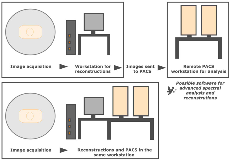
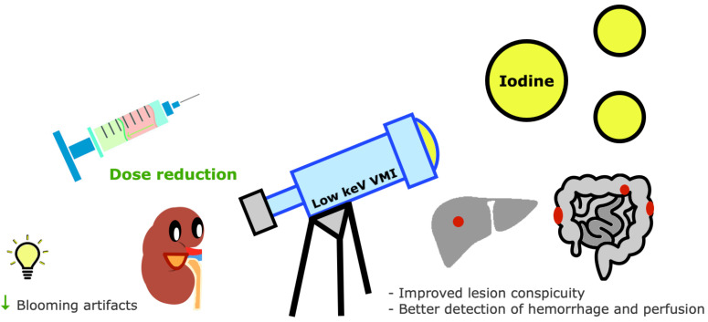
Dual-energy computed tomography (DECT) uses different energy spectrum x-ray beams for differentiating materials with similar attenuation at a certain energy. Compared with single-energy CT, it provides images with better diagnostic performance and a potential reduction of contrast agent and radiation doses. There are different commercially available DECT technologies, with machines that may display two x-ray sources and two detectors, a single source capable of fast switching between two energy levels, a specialized detector capable of acquiring high- and low-energy data sets, and a filter splitting the beam into high- and low-energy beams at the output. Sequential acquisition at different tube voltages is an alternative approach. This narrative review describes the DECT technique using a Q&A format and visual representations. Physical concepts, parameters influencing image quality, postprocessing methods, applicability in daily routine workflow, and radiation considerations are discussed. Differences between scanners are described, regarding design, image quality variabilities, and their advantages and limitations. Additionally, current clinical applications are listed, and future perspectives for spectral CT imaging are addressed. Acknowledging the strengths and weaknesses of different DECT scanners is important, as these could be adapted to each patient, clinical scenario, and financial capability. This technology is undoubtedly valuable and will certainly keep improving.

Keywords: dual-energy CT; dual-layer detector CT; dual-source CT; fast kVp switching; image quality; photon counting; spectral CT; split-filter.


Conflict of interest statement

The authors declare no conflict of interest.

Figures

Figure 1
Figure 1
Flow diagram of the narrative review of the literature.
Figure 2
Figure 2
Schematic representation of the photoelectric effect and Compton scattering. The photoelectric effect is the ejection of an inner K-shell electron (e) of an atom consequent to the interaction of an incident photon (Ph), with subsequent filling of the void by an adjacent shell electron (curved arrow). Compton scattering is the ejection of an outer shell electron (e) of an atom by an incident photon (Ph), resulting in photon scattering with some energy reduction.
Figure 3
Figure 3
Schematic representation of different overlap between low- and high-energy x-ray spectrum. Less overlap (i.e., greater spectral separation) allows better imaging quality.
Figure 4
Figure 4
Postprocessing in DECT imaging. Postprocessing may be performed before (raw data or projection-space domain) or after (image-space domain) the reconstruction of high- or low-energy images. The former has reduced beam-hardening artifacts but requires high computational power. Material decomposition algorithms generate material- and energy-selective images. Iodine may be subtracted from material-specific images, generating a virtual non-contrast image (VNC), or color-coded, creating iodine maps. Virtual monochromatic images (VMIs) mimic single-energy scans. Mixed images are mostly used for routine diagnostic interpretation.
Figure 5
Figure 5
Dual-source DECT. Schematic representation of dual-source DECT scanner and its drawbacks (listed on the left) and strengths (listed on the right). These scanners (commercialized by Siemens Healthineers) are composed of two source-detector pairs dispersed almost perpendicularly. Simultaneous scans are obtained with high- and low-energy spectra (typically 80–100 kVp and 140–150 kVp). The added metallic tin (Sn) filter in the high-energy x-ray tube improves spectral separation, increasing the dose efficiency and CNR (contrast-to-noise ratio), and reduces beam hardening effects. D1—first detector 1, D2—second detector 2, e—energy, FOV—field of view, kVp—peak kilovoltage, ms—miliseconds, s—seconds, S1—first source 1, S2—second source, TR—time resolution.
Figure 6
Figure 6
Single-source DECT with fast kVp switching of tube potential. Schematic representation of a fast kVp switching DECT scanner and its drawbacks (listed on the left) and strengths (listed on the right). These scanners (commercialized by GE Healthcare and Canon Medical Systems) are composed of a specialized generator capable of very fast switching between low- and high-energy spectra projections, which are collected separately by a specialized detector capable of fast sampling. Alternating acquisitions at 80 and 135 or 140 kVp are obtained on each rotation, with a small offset (<0.5°). Aquilion ONE Prism (Canon) offers 16 cm of longitudinal coverage and spectral deep learning reconstruction. e—energy, FOV—field of view, kVp—peak kilovoltage, s—seconds, TR—time resolution.
Figure 7
Figure 7
Dual-layer detector DECT. Schematic representation of a dual-layer detector DECT scanner and its drawbacks (listed on the left) and strengths (listed on the right). These scanners (commercialized by Philips Healthcare) are composed of a single source (fixed energy of 120 or 140 kVp) and a single detector with 2 layers (lower-energy photons preferably absorbed by the top layer and the bottom layer absorbs the remaining higher-energy photons). DE—dual-energy, e—energy, FOV—field of view, kVp—peak kilovoltage, s—seconds.
Figure 8
Figure 8
Single-source split-filter DECT. Schematic representation of single-source split-filter DECT scanner and its drawbacks (listed on the left) and strengths (listed on the right). These scanners (commercialized by Siemens Healthineers) are composed of a single source and detector, with a split-filter of gold (Au) and tin (Sn) at the x-ray tube, respectively, filtering the low- and high-energy beams. Each half of the split beam is captured at corresponding halves of the detector. FOV—field of view, kVp—peak kilovoltage, TR—time resolution.
Figure 9
Figure 9
Single-source DECT with sequential acquisitions. Schematic representation of single-source DECT with sequential acquisitions and its drawbacks (listed on the left) and strengths (listed on the right). These scanners (commercialized by Siemens Healthineers and Canon Medical Systems) are composed of a single source and detector (optional filter). The low-energy scan typically uses 80 kVp, whereas the high-energy scan may use 130 or 140 kVp. The significant time delay between acquisitions leads to poor time resolution (TR) and spectral delay. FOV—field of view, kVp—peak kilovoltage, s—second.
Figure 10
Figure 10
Different DECT workflow algorithms. Necessary reconstructions may be manually generated after the acquisition and the images sent to the picture archiving and communication system (PACS) for remote analysis at a different station (top representation), which may be complemented with advanced software that allows spectral CT analysis and reconstructions. alternatively, most vendors provide lighter workstation versions in the same computer as PACS (bottom representation), requiring a high level of vendor-specific scanner knowledge.
Figure 11
Figure 11
Variable temporal misregistration among DECT scanners. Consecutive scanning techniques are more prone to temporal misregistration and motion. Split-filter DECT scanners require an entire spiral acquisition, so the pitch values are low, resulting in relatively poor temporal registration. Although dual-source DECT scanners have an orthogonal offset of the 2 source-detector pairs inducing spectral delay, its tube design largely improves the temporal resolution. Temporal misregistration is negligible with rapid kVp switching and dual-layer DECT scanners.
Figure 12
Figure 12
Impact of patient size in DECT image quality. Larger bodies may increase the image noise and reduce the image quality (given the insufficient number of photons reaching the detectors, represented by yellow lines). Beam hardening artifacts are more pronounced in larger patient sizes. Potential strategies to improve image quality include increasing the tube current and using noise reduction techniques. Noise is also reduced with the novel photon-counting scanners. The limited field of view (FOV) for dual-energy imaging in dual-source scanners may be a problem in larger patients.
Figure 13
Figure 13
Contrast enhancement improvement with DECT imaging. Iodine density is amplified on low-keV virtual monochromatic images (VMIs). The improved sensitivity for iodine with DECT allows a reduction of iodine dose, which is useful in patients with compromised renal function and reduces blooming artifacts. Other advantages include improved lesion conspicuity and better detection of hemorrhage and perfusion assessment.
Figure 14
Figure 14
Main clinical applications of DECT. Dual-energy CT provides several advantages, including the potential for better lesion depiction and characterization, radiation dose reduction by generating virtual non-contrast (VNC) images, iodine dose reduction, and beam-hardening artifacts reduction. Its benefits are also well-known in radiation oncology. Examples of the many useful applications that have been reported across all body systems are listed here.

Similar articles

Cited by

References

    1. McCollough C.H., Leng S., Yu L., Fletcher J.G. Dual- and Multi-Energy CT: Principles, Technical Approaches, and Clinical Applications. Radiology. 2015;276:637–653. doi: 10.1148/radiol.2015142631. - DOI - PMC - PubMed
    1. Agostini A., Borgheresi A., Mari A., Floridi C., Bruno F., Carotti M., Schicchi N., Barile A., Maggi S., Giovagnoni A. Dual-energy CT: Theoretical principles and clinical applications. La Radiol. Med. 2019;124:1281–1295. doi: 10.1007/s11547-019-01107-8. - DOI - PubMed
    1. Tatsugami F., Higaki T., Nakamura Y., Honda Y., Awai K. Dual-energy CT: Minimal essentials for radiologists. Jpn. J. Radiol. 2022;40:547–559. doi: 10.1007/s11604-021-01233-2. - DOI - PMC - PubMed
    1. Sodickson A.D., Keraliya A., Czakowski B., Primak A., Wortman J., Uyeda J.W. Dual energy CT in clinical routine: How it works and how it adds value. Emerg. Radiol. 2020;28:103–117. doi: 10.1007/s10140-020-01785-2. - DOI - PubMed
    1. Forghani R., De Man B., Gupta R. Dual-Energy Computed Tomography: Physical Principles, Approaches to Scanning, Usage, and Implementation: Part 1. Neuroimaging Clin. N. Am. 2017;27:371–384. doi: 10.1016/j.nic.2017.03.002. - DOI - PubMed