Case Reports
. 2023 Feb 16;9(1):413-419.
doi: 10.3390/tomography9010033.

Trauma to the Eye: Diffusion Restriction on MRI as a Surrogate Marker for Blindness

Affiliations
Case Reports

Trauma to the Eye: Diffusion Restriction on MRI as a Surrogate Marker for Blindness

Andreas Stahl et al. Tomography. .

Abstract

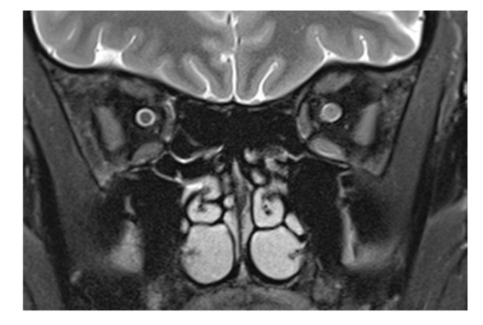
Traumatic optic nerve injury may lead to almost instantaneous blindness. We describe a case of sight loss after a perforating injury to the eye. The case is unusual in that the patient remained conscious and the trauma to the eye was isolated. A full ophthalmological examination was therefore possible within hours as well as early magnetic resonance imaging of the facial skull. High-quality T1-weighted, T2-weighted, and diffusion-weighted imaging could be acquired. The latter included apparent diffusion coefficient maps. There was a loss of the subarachnoid space of the optic nerve, fluid in the retrobulbar fat of the affected eye, and signal changes in the optic nerve. Previous work has been contradictory on the signal of the optic nerve on apparent diffusion coefficient maps in sight loss, with an increase seen by one group and a decrease seen by another. Signal loss on the apparent diffusion coefficient map was seen in the case described here. Signal loss on apparent diffusion coefficient maps may thus be used as a surrogate marker of sight loss in patients who are unconscious or otherwise unable to cooperate in ophthalmological exams.

Keywords: MRI; diffusion-weighted imaging; optic nerve; sight loss; traumatic brain injury.


Conflict of interest statement

The authors declare no conflict of interest.

Figures

Figure 1
Figure 1
Examination of the right eye revealed a superficial conjunctival wound nasally and a dilated pupil (a). On the MRI scan, there is loss of the subarachnoid space in the right optic nerve (arrows in panel (b): the subarachnoid space of the left optic nerve is normal). The respective transverse T2-weighted image shows a central signal increase in the optic nerve (arrow in (c)). On the diffusion-weighted imaging, a punctuate signal increase in the optic nerve in the same location is seen (arrow in (d)); the corresponding ADC map shows signal loss in the same location (arrow in (e)). The globe as well as the left eye and orbit are unchanged, while there is a diffuse signal increase (arrows) in the right retrobulbar space nasally (f,g).
Figure 1
Figure 1
Examination of the right eye revealed a superficial conjunctival wound nasally and a dilated pupil (a). On the MRI scan, there is loss of the subarachnoid space in the right optic nerve (arrows in panel (b): the subarachnoid space of the left optic nerve is normal). The respective transverse T2-weighted image shows a central signal increase in the optic nerve (arrow in (c)). On the diffusion-weighted imaging, a punctuate signal increase in the optic nerve in the same location is seen (arrow in (d)); the corresponding ADC map shows signal loss in the same location (arrow in (e)). The globe as well as the left eye and orbit are unchanged, while there is a diffuse signal increase (arrows) in the right retrobulbar space nasally (f,g).
Figure 2
Figure 2
T2-weighted frontal image in a multiple sclerosis patient with optic neuritis. While the subarachnoid space has a normal diameter on the left, an enlarged optic nerve encroaches in the same space, recognizable by the smaller liquor-filled rim of the nerve. Leakage of cerebrospinal liquor after direct laceration of the sheath, however, cannot be counted out. The diffuse signal increase of the nasal retrobulbar space in Figure 1f,g may be explained by cerebrospinal liquor leaking from an injured optic nerve sheath and by edema.

References

    1. Ramsay J.H. Optic nerve injury in fracture of the canal. Br. J. Ophthalmol. 1979;63:607–610. doi: 10.1136/bjo.63.9.607. - DOI - PMC - PubMed
    1. Seitz R. Etiology and genesis of acute blindness following blunt trauma to the head. Klin. Monatsbl. Augenheilkd. 1963;143:414–429. - PubMed
    1. Hughes B. Indirect injury of optic nerves and chiasma. Bull. Johns Hopkins Hospit. 1962;111:98–126. - PubMed
    1. Steinsapir K.D., Goldberg R.A. Traumatic optic neuropathy. Surv. Ophthalmol. 1994;38:487–518. doi: 10.1016/0039-6257(94)90145-7. - DOI - PubMed
    1. Goelz L., Syperek A., Heske S., Mutze S., Hosten N., Kirsch M. Retrospective Cohort Study of Frequency and Patterns of Orbital Injuries on Whole-Body CT with Maxillofacial Multi-Slice CT. Tomography. 2021;7:373–386. doi: 10.3390/tomography7030033. - DOI - PMC - PubMed

Publication types