Review
. 2023 Apr;44(4):222-236.
doi: 10.1016/j.tips.2023.01.003. Epub 2023 Feb 23.

Metastatic colorectal cancer: mechanisms and emerging therapeutics

Affiliations
Review

Metastatic colorectal cancer: mechanisms and emerging therapeutics

Alice E Shin et al. Trends Pharmacol Sci. 2023 Apr.

Abstract

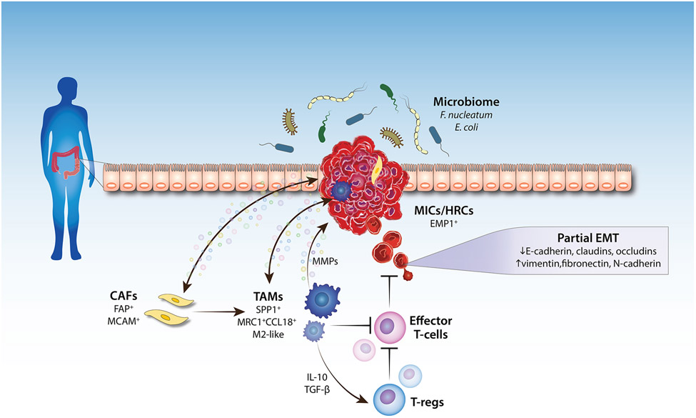
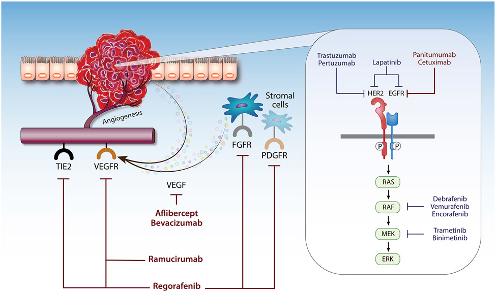
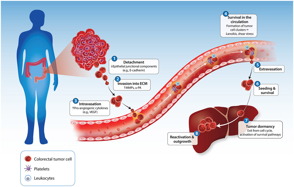
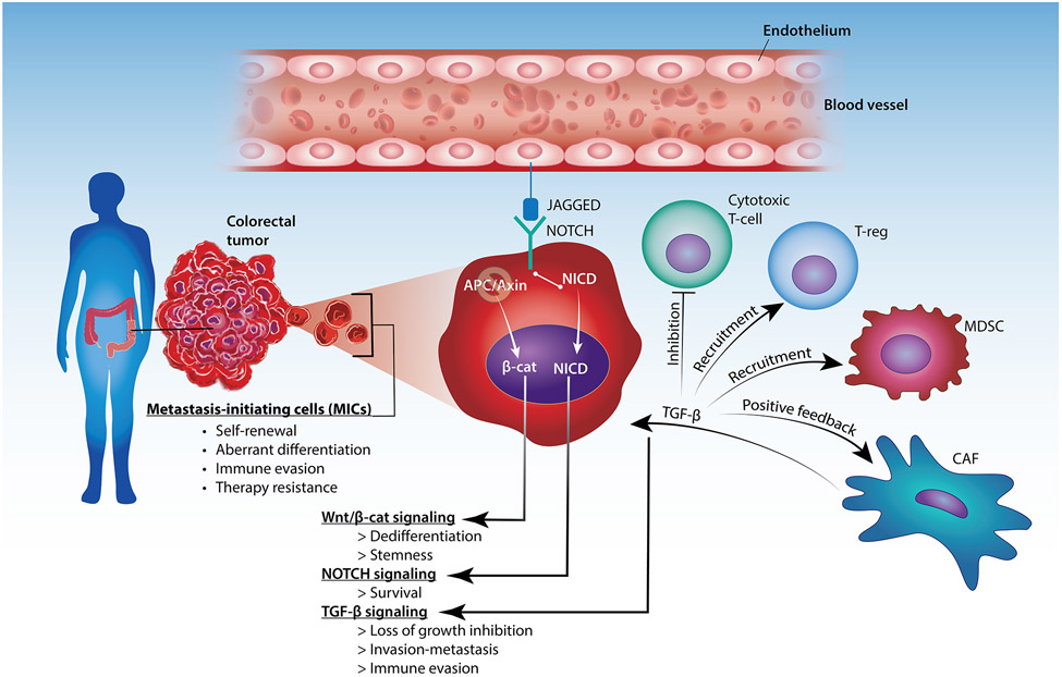
Metastatic colorectal cancer (mCRC) remains a lethal disease with an approximately 14% 5-year survival rate. While early-stage colorectal cancer (CRC) can be cured by surgery with or without adjuvant chemotherapy, mCRC cannot be eradicated due to a large burden of disseminated cancer cells comprising therapy-resistant metastasis-competent cells. To address this gap, recent studies have focused on further elucidating the molecular mechanisms underlying colorectal metastasis and recognizing the limitations of available therapeutic interventions. In this review, we discuss newfound factors that regulate CRC cell dissemination and colonization of distant organs, such as genetic mutations, identification of metastasis-initiating cells (MICs), epithelial-mesenchymal transition (EMT), and the tumor microenvironment (TME). We also review current treatments for mCRC, therapeutic regimens undergoing clinical trials, and trending preclinical studies being investigated to target treatment-resistant mCRC.

Keywords: cancer therapeutics; colorectal cancer; immunotherapy; metastasis; targeted therapy.


Conflict of interest statement

Declaration of interests The authors declare no interests.

Figures

Figure 1.
Figure 1.. Factors that affect colorectal cancer metastasis.
The tumor microenvironment (TME) consists of tumor cells, resident host cells (the colonic epithelium), immune cells, endothelial cells, neurons, adipocytes, secreted factors, and the extracellular matrix (ECM). The interplay between the cells within the TME and the tumor cells, as well as the interplay between the gut microbiome and the tumor cells, regulate tumor invasion and metastasis (namely, intravasation into blood vessels). Changes in the expression of epithelial-mesenchymal transition (EMT) genes occur within the subset of tumor cells that acquire metastatic properties. Abbreviations: MIC, metastasis-initiating cell; HRC, high-relapse cell; EMP1, epithelial membrane protein 1; MMP, matrix metalloproteinase; CAF, cancer-associated fibroblast; TAM, tumor-associated macrophage; T-reg, regulatory T-cell; TGF-β, transforming growth factor-β. Arrows pointing to the tumor indicate tumor-promoting effect.
Figure 2.
Figure 2.. Targeted therapy for metastatic colorectal cancer.
Diagram illustrating the mechanism by which biological agents target the tumor cells or the tumor microenvironment (TME) to inhibit CRC metastasis. The EGFR encompasses four closely related receptor tyrosine kinases (i.e., EGFR, HER2-4). Dimerization of the receptors leads to their phosphorylation and subsequent activation of the effector proteins of the MAPK pathway (RAS, RAF, MEK, and ERK). Trastuzumab, pertuzumab, lapatinib, panitumumab, cetuximab, dabrafenib, vemurafenib, encorafenib, trametinib, and binimetinib target the HER2/EGFR-mediated MAPK pathway. Drugs such as bevacizumab, aflibercept, ramucirumab, and regorafenib target angiogenesis by inhibiting the VEGF receptors expressed on endothelial cells. Names of drugs in red indicate US FDA-approved therapy. Names of drugs in blue indicate drugs in clinical trials. Abbreviations: HER2, human epidermal growth factor receptor 2; EGFR, epidermal growth factor receptor; VEGF, vascular endothelial growth factor; VEGFR, VEGF receptor; FGFR, fibroblast growth factor receptor; PDGFR, platelet-derived growth factor receptor; TIE2, tunica interna endothelial cell kinase 2.
Figure I.
Figure I.. The invasion-metastasis cascade in colorectal cancer.
Step 1: tumor cells detach from one another. Step 2: tumor cells invade into the ECM by degrading the basement membrane. Step 3: tumor cells induce angiogenesis and enter the circulation. Step 4: tumor cells survive within the circulation by clustering with each other, leukocytes, and platelets. Step 5: tumor cells that have survived the circulation exit the bloodstream into the stroma of the target organ. Step 6: tumor cells seed into the secondary site. Step 7: tumor cells enter a dormant state, characterized by exit from the cell cycle and activation of survival pathways. Step 8: tumor cells are reactivated, allowing for their outgrowth and colonization at the secondary site. Abbreviations: MMP, matrix metalloproteinase; u-PA, urokinase plasminogen activator; VEGF, vascular endothelial growth factor.
Figure II.
Figure II.. Metastasis-initiating cells (MICs) in colorectal cancer.
Metastases arise from a subset of tumor cells that have acquired invasiveness due to the activation of the NOTCH, Wnt/β-catenin, and TGF-β signaling pathways. Interaction between JAGGED (expressed by endothelial cells) and NOTCH (expressed by the tumor cells) allow for survival of the tumor cells around tumor vessels. Loss of APC or Axin promotes Wnt/β-catenin signaling and induce dedifferentiation and stemness of the MICs. MICs are resistant to the cytostatic effects of TGF-β signaling, which can also create an immune-suppressive microenvironment and further promote invasion of MICs. Abbreviations: TGF-β, transforming growth factor-β; β-cat, β-catenin; NICD, Notch intracellular domain; APC, adenomatous polyposis coli; T-reg, regulatory T-cell; MDSC, myeloid-derived suppressor cell; CAF, cancer-associated fibroblast.

References

    1. Rumpold H et al. (2020) Prediction of mortality in metastatic colorectal cancer in a real-life population: a multicenter explorative analysis. BMC Cancer 20, 1149. 10.1186/s12885-020-07656-w - DOI - PMC - PubMed
    1. Xie YH et al. (2020) Comprehensive review of targeted therapy for colorectal cancer. Signal Transduct Target Ther 5, 22. 10.1038/s41392-020-0116-z - DOI - PMC - PubMed
    1. Ganesh K et al. (2019) Immunotherapy in colorectal cancer: rationale, challenges and potential. Nat Rev Gastroenterol Hepatol 16, 361–375. 10.1038/s41575-019-0126-x - DOI - PMC - PubMed
    1. Nagao K et al. (2022) The Complete Loss of p53 Expression Uniquely Predicts Worse Prognosis in Colorectal Cancer. Int J Mol Sci 23. 10.3390/ijms23063252 - DOI - PMC - PubMed
    1. Hassin O et al. (2022) Different hotspot p53 mutants exert distinct phenotypes and predict outcome of colorectal cancer patients. Nat Commun 13, 2800. 10.1038/s41467-022-30481-7 - DOI - PMC - PubMed

Publication types

MeSH terms