. 2023 Feb 24;14(1):1061.
doi: 10.1038/s41467-023-36620-y.

Citrullination modulates antigen processing and presentation by revealing cryptic epitopes in rheumatoid arthritis

Affiliations

Citrullination modulates antigen processing and presentation by revealing cryptic epitopes in rheumatoid arthritis

Ashley M Curran et al. Nat Commun. .

Abstract

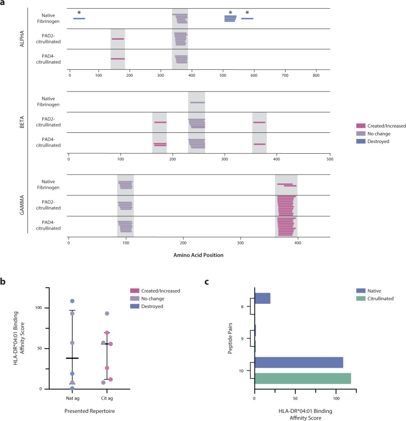
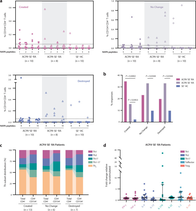
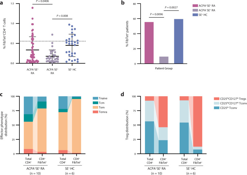
Cryptic peptides, hidden from the immune system under physiologic conditions, are revealed by changes to MHC class II processing and hypothesized to drive the loss of immune tolerance to self-antigens in autoimmunity. Rheumatoid arthritis (RA) is an autoimmune disease characterized by immune responses to citrullinated self-antigens, in which arginine residues are converted to citrullines. Here, we investigate the hypothesis that citrullination exposes cryptic peptides by modifying protein structure and proteolytic cleavage. We show that citrullination alters processing and presentation of autoantigens, resulting in the generation of a unique citrullination-dependent repertoire composed primarily of native sequences. This repertoire stimulates T cells from RA patients with anti-citrullinated protein antibodies more robustly than controls. The generation of this unique repertoire is achieved through altered protease cleavage and protein destabilization, rather than direct presentation of citrulline-containing epitopes, suggesting a novel paradigm for the role of protein citrullination in the breach of immune tolerance in RA.


Conflict of interest statement

The authors declare the following competing interests: E.D. is an inventor on a licensed patent (US patent no. 8,975,033) and licensed provisional patent (US patent no. 62/481,158) related to the use of antibodies to PAD3 and PAD2, respectively, in identifying clinically informative disease subsets in RA, and has received consulting fees from Celgene and Bristol Myers Squibb and research support from Pfizer, Celgene, and Bristol Myers Squibb outside of this work. The remaining authors declare no competing interests.

Figures

Fig. 1
Fig. 1. Citrullination alters antigen processing, resulting in the simultaneous generation of cryptic peptides and the destruction of previously dominant peptides.
a Log2 enrichment of peptides from proteolytic mapping with cathepsins BSH across the primary amino acid sequence of each protein. Amino acid positions with positive values are enriched in the PAD2- or PAD4-citrullinated (cit) samples compared to the native (nat) samples, while those with negative values are reduced by citrullination. Citrulline residues are denoted by the filled blue circles on x axes. b Volcano plots representing peptides with significantly altered abundance. Vertical lines denote a fold change of 2, and the horizontal line denotes a cutoff of 0.05 for the FDR-corrected P value (q value), calculated by paired two-sided Student’s t tests. Peptides to the left or right of vertical dashed lines and above horizontal dashed line are deemed to be significantly different between the groups. Proteolytic mapping was performed in replicates of four (fibrinogen and vimentin) or six (hnRNP A2/B1).
Fig. 2
Fig. 2. Citrullination induces proximal and distal changes in processing relative to citrullinated sites.
a Percentage of significantly created and destroyed regions derived from PAD2- and PAD4-citrullinated antigens that contain citrulline residues. b Frequency of citrulline residues in each position (P4-P4’) of citrulline-containing cleavage sites enriched in the citrullinated antigens. P values were calculated via bootstrapping to compare the observed frequency of citrullines to the expected frequency of citrullines in each position. Only significant P values (≤0.05) are shown. c Violin plots depicting the actual minus expected distance from each significantly changed region to the nearest citrulline residue in the linear amino acid sequence. d Arginine frequency shown for all autoantigens used in this study. The horizontal dotted line represents the average vertebrate protein arginine content. e Antigen processing change score versus % arginine content (frequency) for each PAD2- or PAD4-citrullinated (cit) antigen.
Fig. 3
Fig. 3. Citrullination impacts protein dynamics by promoting the destabilization of protein folding.
a PyMOL structural alignment of native (gray) and PAD2/4-combined citrullinated (colored by antigen) PDB structures predicted by AlphaFold AI system, excluding fibrinogen α chain due to computational constraints. Mass spectrometry-confirmed citrullination sites from PAD2 and PAD4 combined are denoted as purple spheres on the citrullinated structure. b Predicted PAD2-citrullinated structures with significantly changed regions color-coded according to legend and PAD2 citrullination sites denoted as purple spheres. See Supplementary Fig. 4 for PAD4-citrullinated structures. c Violin plots depicting the actual minus expected distance from each significantly changed region to the nearest citrulline residue in the 3-dimensional structure. d, e Antigen processing change score versus RMSD value or 1/TM-score, respectively, analyzed by simple linear regression for PAD2- or PAD4-citrullinated (cit) vimentin, fibrinogen β, and hnRNP A2/B1.
Fig. 4
Fig. 4. Putative RA-associated HLA-DR peptide-binding cores primarily consist of native sequences enriched by citrullination.
ac Log2 enrichment of putative binding cores (<500 nM) predicted by the NetMHCII-2.3 algorithm, denoted by the first three amino acids of the peptide core. Cores with positive values are enriched in the PAD2- or PAD4-citrullinated (cit) samples, while those with negative values are reduced by citrullination. *Denotes a uniquely created or destroyed peptide, and color-coded cores are those that contained a citrullination site. d Pie charts representing the proportion of all putative binding cores belonging to several categories (given by legend) denoting the behavior of each core in response to citrullination.
Fig. 5
Fig. 5. Citrullination alters the naturally presented peptide repertoire presented by human mo-DCs.
a NAPA performed on mo-DCs from a SE+ healthy donor pulsed with native and citrullinated fibrinogen. Peptides from the α, β, and γ chains of fibrinogen are shown. Each colored line represents a unique peptide coded according to the legend, and gray bars denote peptides with identical core motifs between samples. *Denotes a citrullination site. b HLA-DRB1*04:01 relative binding affinity scores for the presented peptide repertoires derived from the native antigen (Nat ag; n = 6 peptides) and the citrullinated antigen (Cit ag; n = 7 peptides) were measured by the ProImmune REVEAL® assay, which represents a surrogate for binding affinity through the quantification of MHC stabilization relative to an internal positive control peptide. The center lines represent the median, and the error bars denote the interquartile range. c HLA-DRB1*04:01 relative binding affinity scores as measured by the ProImmune REVEAL® assay for the three native and citrullinated peptide pairs.
Fig. 6
Fig. 6. Unmodified, citrullination-dependent epitopes stimulate ACPA+SE+ RA patient CD4+ T cells more robustly than native antigen–derived epitopes.
a PBMCs from ACPA+SE+ RA patients (n = 10), ACPASE+RA patients (n = 8), and SE+ healthy controls (n = 10) were stimulated with created peptides (1–4), no change peptides (5–7), and destroyed peptides (8–10) derived from NAPA (Fig. 5a). Percentage of CD154+CD4+ T-cell response to each peptide is shown. b Percentage of potential T-cell responses (where the number of potential T-cell responses were defined as total patients per group multiplied by the total peptides per group) is shown. Two-sided χ2 tests were used to compare the frequency of T-cell responses between patient groups. Only significant P values (≤0.05) are shown. c Distribution of ACPA+SE+ RA patient CD4+ T-helper subsets (denoted in legend) in either the total CD4+ or the CD4+CD154+ T-cell population in response to stimulation with created, no change, or destroyed peptides. d ACPA+SE+ RA patient (n = 18) PBMC cytokine secretion in response to 48-h stimulation with the created peptide pool, normalized to unstimulated samples. The dotted line at a fold change of 1 represents the normalized unstimulated values. Data are presented as median and IQR.
Fig. 7
Fig. 7. ACPA+SE+ RA patient CD4+ T cells specific for citrullination-dependent epitopes tend to be activated effector memory cells.
a MHC class II tetramers containing three created fibrinogen peptides derived from NAPA (Fig. 5a) were used to stain PBMCs from ACPA+SE+ RA patients (n = 18), ACPASE+RA patients (n = 10), and SE+ healthy controls (n = 10). The percentage of fibrinogen tetramer–positive (FibTet+) CD4+ T cells are shown for each patient group, and the dotted line denotes the positivity threshold of 0.5547%. Data are presented as mean ± SD, and P values were calculated by ordinary one-way ANOVA with Tukey’s multiple comparisons test. Only significant P values (≤0.05) are shown. b The percentage of individuals that bound any FibTet is shown for each group. Two-sided χ2 tests were used to compare the frequency of T-cell responses between patient groups. Only significant P values (≤0.05) are shown. c, d Distribution of effector phenotypes (c) and regulatory T cells (d) for ACPA+SE+ RA patient and SE+ healthy control fibrinogen tetramer–positive T cells. Phenotypes denoted in key.

Comment in

Similar articles

Cited by

References

    1. Sercarz EE, Maverakis E. MHC-guided processing: binding of large antigen fragments. Nat. Rev. Immunol. 2003;3:621–629. doi: 10.1038/nri1149. - DOI - PubMed
    1. Watts C, Matthews SP, Mazzeo D, Manoury B, Moss CX. Asparaginyl endopeptidase: case history of a class II MHC compartment protease. Immunol. Rev. 2005;207:218–228. doi: 10.1111/j.0105-2896.2005.00312.x. - DOI - PubMed
    1. Manoury B, et al. An asparaginyl endopeptidase processes a microbial antigen for class II MHC presentation. Nature. 1998;396:695–699. doi: 10.1038/25379. - DOI - PubMed
    1. Antoniou AN, Blackwood S-L, Mazzeo D, Watts C. Control of antigen presentation by a single protease cleavage site. Immunity. 2000;12:391–398. doi: 10.1016/S1074-7613(00)80191-0. - DOI - PubMed
    1. Darrah E, et al. Proteolysis by granzyme B enhances presentation of autoantigenic peptidylarginine deiminase 4 epitopes in rheumatoid arthritis. J. Proteome Res. 2017;16:355–365. doi: 10.1021/acs.jproteome.6b00617. - DOI - PMC - PubMed

Publication types